

Shizuo Akira
Immune Response and Inflammation
The University of Osaka
About
Research focus
Immune Response and Inflammation
Impact
1,481 pubs · 335,816 cites · h-index 269
Affiliations
Publications
preclinicalCirculation Highlight
NOV 2004KnuefermannCirculation
TLR2 deficiency protects against contractile dysfunction and blunts inflammation in Staph aureus cardiac challenge.
TLR2 signaling mediates myocardial contractile dysfunction and cytokine production during Staphylococcus aureus sepsis in a murine model.
Original Research
JUL 2025Furlong-Silva
Mgl2⁺ cDC2 triggering of fungal allergic inflammation depends on a spore induced glycolytic shift fuelled by local availability of glucose
Research reveals that metabolic changes in dendritic cells impact allergic inflammation in mice, indicating glucose's key role.
Prognostic Study
JUL 2025NiitsuThe Journal of Pathology
Spatial transcriptomics identifies SPARC as a prognostic marker in interstitial lung diseases
Interstitial lung diseases (ILDs) encompass a diverse group of pulmonary disorders, with progressive fibrosis leading to poor prognosis. Here we aimed to identify key molecules involved in progressive fibrosis across various ILDs, using spatial transcriptomics (ST). ST analysis (Visium) was performed on lung cryobiopsy specimens from five patients with various ILDs. Two cases, rich in young fibrotic lesions, as defined by fibroblastic foci and destructive alveolar organization, were selected for spatial high-dimensional weighted gene coexpression network analysis (hdWGCNA) to identify key gene networks with biological significance in active fibrosis. We utilized public single-cell RNA sequencing datasets of various ILDs, performed enrichment analysis and trajectory-based differential expression analysis, and quantified cell-cell communication to evaluate the involvement of the spatially extracted module in fibrosis. Immunohistochemical staining of the extracted molecules was performed. Using hdWGCNA, we identified a distinct gene module (the SM2 module) enriched in young fibrotic lesions. The SM2 module was characterized by distinct features of fibroblast activation that were represented across various lesions. Key hub genes within this module, including COL1A2, COL3A1, COL1A1, and SPARC, formed a robust coexpression network. Immunohistochemical staining showed that SPARC, a component of the SM2 module, was highly expressed in young fibrotic lesions, but not in old scarring lesions, across various ILDs. To assess the prognostic significance of SPARC immunohistochemical expression, we extended our analysis to a cohort of 71 patients with unclassifiable ILDs (uILDs), a particularly heterogeneous subtype with unclear pathogenesis and limited treatment options. Higher SPARC levels in the upper, lower, or both lung lobes in uILD were significantly associated with poor overall survival. In summary, an integrated cross-disease approach using ST revealed key gene expression patterns central to active fibrosis and successfully identified SPARC as a potentially beneficial prognostic marker.
Original Research
MAR 2025PoplieLife
The autophagy protein, ATG14 safeguards against unscheduled pyroptosis activation to enable embryo transport during early pregnancy
Recurrent pregnancy loss (RPL), characterized by two or more failed clinical pregnancies, poses a significant challenge to reproductive health. In addition to embryo quality and endometrial function, proper oviduct function is also essential for successful pregnancy establishment. Therefore, structural abnormalities or inflammation resulting from infection in the oviduct may impede the transport of embryos to the endometrium, thereby increasing the risk of miscarriage. However, our understanding of the biological processes that preserve the oviductal cellular structure and functional integrity is limited. Here, we report that autophagy-related protein ATG14 plays a crucial role in maintaining the cellular integrity of the oviduct by controlling inflammatory responses, thereby supporting efficient embryo transport. Specifically, the conditional depletion of the autophagy-related gene, Atg14 in the oviduct causes severe structural abnormalities compromising its cellular integrity leading to the abnormal retention of embryos. Interestingly, the selective loss of Atg14 in oviduct ciliary epithelial cells did not impact female fertility, highlighting the specificity of ATG14 function in distinct cell types within the oviduct. Mechanistically, loss of Atg14 triggered unscheduled pyroptosis via altering the mitochondrial integrity leading to inappropriate embryo retention and impeded embryo transport in the oviduct. Finally, pharmacological activation of pyroptosis in pregnant mice phenocopied the genetically induced defect and caused impairment in embryo transport. Together, we found that ATG14 safeguards against unscheduled pyroptosis activation to enable embryo transport from the oviduct to uterus for the successful implantation. Of clinical significance, these findings provide possible insights into the underlying mechanism(s) of early pregnancy loss and might aid in developing novel prevention strategies using autophagy modulators.
Original Research
NOV 2024SakaguchiNutrients
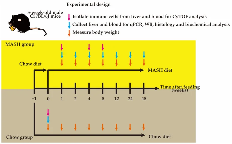
Novel Choline-Deficient and 0.1%-Methionine-Added High-Fat Diet Induces Burned-Out Metabolic-Dysfunction-Associated Steatohepatitis with Inflammation by Rapid Immune Cell Infiltration on Male Mice
Novel MASLD model data hypothesis-generating; leaves open whether it improves drug development success.
Original Research
NOV 2024PoplieLife
The autophagy protein, ATG14 safeguards against unscheduled pyroptosis activation to enable embryo transport during early pregnancy
Recurrent pregnancy loss (RPL), characterized by two or more failed clinical pregnancies, poses a significant challenge to reproductive health. In addition to embryo quality and endometrial function, proper oviduct function is also essential for successful pregnancy establishment. Therefore, structural abnormalities or inflammation resulting from infection in the oviduct may impede the transport of embryos to the endometrium, thereby increasing the risk of miscarriage. However, our understanding of the biological processes that preserve the oviductal cellular structure and functional integrity is limited. Here, we report that Atg14-dependent autophagy plays a crucial role in maintaining the cellular integrity of the oviduct by controlling inflammatory responses, thereby supporting efficient embryo transport. Specifically, the conditional depletion of the autophagy-related gene, Atg14 in the oviduct causes severe structural abnormalities compromising its cellular integrity leading to the abnormal retention of embryos. Interestingly, the selective loss of Atg14 in oviduct ciliary epithelial cells did not impact female fertility, highlighting the specificity of ATG14 function in distinct cell types within the oviduct. Mechanistically, loss of Atg14 triggered unscheduled pyroptosis via altering the mitochondrial integrity leading to inappropriate embryo retention and impeded embryo transport in the oviduct. Finally, pharmacological activation of pyroptosis in pregnant mice phenocopied the genetically induced defect and caused impairment in embryo transport. Together, we found that ATG14 safeguards against unscheduled pyroptosis activation to enable embryo transport from the oviduct to uterus for the successful implantation. Of clinical significance, these findings provide possible insights into the underlying mechanism(s) of early pregnancy loss and might aid in developing novel prevention strategies using autophagy modulators.
Retrospective Study
OCT 2024FukushimaAntimicrobial Agents and Chemotherapy
Variability of macrolide-resistant profile in Mycobacterium avium complex pulmonary disease
This single-center retrospective study aimed to analyze the variability of macrolide resistance (MR) in 68 patients with Mycobacterium avium complex pulmonary disease. Among 25 patients treated without macrolides, 13 (52%) reverted to macrolide-susceptible (MS) profiles. Only one (2%) of 43 patients who continued macrolide treatment showed this change. We compared 30 MR isolates with recent specimens. Among them, seven shifted to MS (five attributed to clonally related strains; two resulting from reinfection or polyclonal infection).
Original Research
SEP 2024NayerNature Communications
Local administration of regulatory T cells promotes tissue healing
Regulatory T cells (Tregs) are crucial immune cells for tissue repair and regeneration. However, their potential as a cell-based regenerative therapy is not yet fully understood. Here, we show that local delivery of exogenous Tregs into injured mouse bone, muscle, and skin greatly enhances tissue healing. Mechanistically, exogenous Tregs rapidly adopt an injury-specific phenotype in response to the damaged tissue microenvironment, upregulating genes involved in immunomodulation and tissue healing. We demonstrate that exogenous Tregs exert their regenerative effect by directly and indirectly modulating monocytes/macrophages (Mo/MΦ) in injured tissues, promoting their switch to an anti-inflammatory and pro-healing state via factors such as interleukin (IL)-10. Validating the key role of IL-10 in exogenous Treg-mediated repair and regeneration, the pro-healing capacity of these cells is lost when Il10 is knocked out. Additionally, exogenous Tregs reduce neutrophil and cytotoxic T cell accumulation and IFN-γ production in damaged tissues, further dampening the pro-inflammatory Mo/MΦ phenotype. Highlighting the potential of this approach, we demonstrate that allogeneic and human Tregs also promote tissue healing. Together, this study establishes exogenous Tregs as a possible universal cell-based therapy for regenerative medicine and provides key mechanistic insights that could be harnessed to develop immune cell-based therapies to enhance tissue healing. Regulatory T cells (Tregs) are known for suppressing inflammatory processes, but their full capacity for tissue regeneration is yet to be harnessed. Here, the authors demonstrate the efficiency of Tregs in facilitating tissue healing in mouse models of bone, muscle, and skin injury, with monocytes/macrophages and interleukin-10 playing a key mechanistic role in the process.
Blinded Clinical Trial
SEP 2024TanakaRMD Open
Safety, pharmacokinetics, biomarker response and efficacy of E6742: a dual antagonist of Toll-like receptors 7 and 8, in a first in patient, randomised, double-blind, phase I/II study in systemic lupus erythematosus
Objectives To evaluate the safety, tolerability, pharmacokinetics (PK), biomarker response and efficacy of E6742 in a phase I/II study in patients with systemic lupus erythematosus (SLE). Methods Two sequential cohorts of patients with SLE were enrolled and randomised to 12 weeks of two times per day treatment with E6742 (100 or 200 mg; n=8 or 9) or placebo (n=9). The primary endpoint was safety, the secondary endpoints were PK and interferon gene signature (IGS), and the exploratory endpoints were efficacy and biomarker. Results The proportion of patients with any treatment-emergent adverse events (TEAEs) was 58.8% in the E6742 group (37.5% (3/8 patients) for 100 mg; 77.8% (7/9 patients) for 200 mg) and 66.7% (6/9 patients) in the placebo group. No Common Terminology Criteria for Adverse Events≥Grade 3 TEAEs occurred. PK parameters were similar to these in previous phase I studies in healthy adults. The IGS and levels of proinflammatory cytokines after ex-vivo challenge with a Toll-like receptor 7/8 agonist were immediately decreased by E6742 treatment. The response rate of the British Isles Lupus Assessment Group-based Composite Lupus Assessment at week 12 was 37.5% (3/8 patients) for E6742 100 mg, 57.1% (4/7 patients) for E6742 200 mg and 33.3% (3/9 patients) for placebo group. Conclusions E6742 had a favourable safety profile and was well tolerated, with suppression of IGS responses and preliminary efficacy signals in patients with SLE. These results provide the first clinical evidence to support E6742 in the treatment of SLE, and support larger, longer-term clinical trials. Trial registration number NCT05278663 .
Original Research
JUN 2024HobroChemical & Biomedical Imaging
Correlative Quantitative Raman Chemical Imaging and MCR–ALS in Mouse NASH Model Reveals Direct Relationships between Diet and Resultant Liver Pathology
Raman imaging has the capability to provide unlabeled, spatially aware analysis of chemical components, with no a priori assumptions. Several lifestyle diseases such as nonalcoholic steatohepatitis (NASH) can appear in the liver as changes in the nature, abundance, and distribution of lipids, proteins, and other biomolecules and are detectable by Raman imaging. In order to identify which of these liver-associated changes occur as a direct result of the diet and which are secondary effects, we developed correlative imaging and analysis of diet and liver samples. Oleic acid was found to be a direct contributor to NASH liver composition, whereas protein and collagen distributions were found to be affected in a manner consistent with early fibrotic transformation, as a secondary consequence of the high-fat diet.
Original Research
JUN 2024PopliBaylor College of Medicine
The autophagy protein, ATG14 safeguards against unscheduled pyroptosis activation to enable embryo transport during early pregnancy
Recurrent pregnancy loss (RPL), characterized by two or more failed clinical pregnancies, poses a significant challenge to reproductive health. In addition to embryo quality and endometrial function, proper oviduct function is also essential for successful pregnancy establishment. Therefore, structural abnormalities or inflammation resulting from infection in the oviduct may impede the transport of embryos to the endometrium, thereby increasing the risk of miscarriage. However, our understanding of the biological processes that preserve the oviductal cellular structure and functional integrity is limited. Here, we report that autophagy-related protein ATG14 plays a crucial role in maintaining the cellular integrity of the oviduct by controlling inflammatory responses, thereby supporting efficient embryo transport. Specifically, the conditional depletion of the autophagy-related gene, Atg14 in the oviduct causes severe structural abnormalities compromising its cellular integrity leading to the abnormal retention of embryos. Interestingly, the selective loss of Atg14 in oviduct ciliary epithelial cells did not impact female fertility, highlighting the specificity of ATG14 function in distinct cell types within the oviduct. Mechanistically, loss of Atg14 triggered unscheduled pyroptosis via altering the mitochondrial integrity leading to inappropriate embryo retention and impeded embryo transport in the oviduct. Finally, pharmacological activation of pyroptosis in pregnant mice phenocopied the genetically induced defect and caused impairment in embryo transport. Together, we found that ATG14 safeguards against unscheduled pyroptosis activation to enable embryo transport from the oviduct to uterus for the successful implantation. Of clinical significance, these findings provide possible insights into the underlying mechanism(s) of early pregnancy loss and might aid in developing novel prevention strategies using autophagy modulators.
Original Research
JUN 2024PopliBaylor College of Medicine
The autophagy protein, ATG14 safeguards against unscheduled pyroptosis activation to enable embryo transport during early pregnancy
Recurrent pregnancy loss (RPL), characterized by two or more failed clinical pregnancies, poses a significant challenge to reproductive health. In addition to embryo quality and endometrial function, proper oviduct function is also essential for successful pregnancy establishment. Therefore, structural abnormalities or inflammation resulting from infection in the oviduct may impede the transport of embryos to the endometrium, thereby increasing the risk of miscarriage. However, the precise cellular mechanisms that maintain the structural and functional integrity of the oviduct are not studied yet. Here, we report that autophagy is critical for maintaining the oviduct homeostasis and keeping the inflammation under check to enable embryo transport. Specifically, the loss of the autophagy-related gene, Atg14 in the oviduct causes severe structural abnormalities compromising its cellular plasticity and integrity leading to the retention of embryos. Interestingly, the selective loss of Atg14 in oviduct ciliary epithelial cells did not impact female fertility, highlighting the specificity of ATG14 function in distinct cell types within the oviduct. Mechanistically, loss of Atg14 triggered unscheduled pyroptosis leading to inappropriate embryo retention and impeded embryo transport in the oviduct. Finally, pharmacological activation of pyroptosis in pregnant mice led to an impairment in embryo transport. Together, we found that ATG14 safeguards against unscheduled pyroptosis activation to enable embryo transport from the oviduct to uterus for the successful implantation. Of clinical significance, these findings provide possible insights on the underlying mechanism(s) of early pregnancy loss and might aid in developing novel prevention strategies using autophagy modulators.
Blinded Clinical Trial
JUN 2024TanakaAnnals of the Rheumatic Diseases
Pos0536 Safety, Pharmacokinetics, Biomarker, and Efficacy of E6742, a Dual Antagonist of Toll-Like Receptor 7 and 8, in a First-in-Patient Phase1/2 Study in Systemic Lupus Erythematosus
Background: E6742 is a novel, first-in-class, orally available, small molecule inhibitor of toll-like receptors (TLR) 7 and 8. Based on preclinical evidence, E6742 inhibits the activation of TLR7 and TLR8 which promote autoimmune response in systemic lupus erythematosus (SLE). The safety results from previous Phase 1 studies in healthy adults demonstrated that E6742 has an adequate safety and tolerability profile. Objectives: To evaluate the safety, tolerability, pharmacokinetics (PK), biomarker, and efficacy of E6742 in a Phase 1/2, randomized, double-blind, placebo-controlled study in patients with SLE for the first time (NCT05278663). Methods: SLE patients received 100 mg E6742 or placebo twice daily in cohort 1, and 200 mg E6742 or placebo twice daily in cohort 2. In each cohort, patients were randomized to either E6742 or placebo in a ratio of 2:1 and received multiple oral administrations of E6742 or placebo for 12 weeks. Results: A total of 12 patients (8 for E6742 100 mg, 4 for placebo) were enrolled in the cohort 1 and subsequently 14 patients (9 for E6742 200 mg, 5 for placebo) were enrolled in the cohort 2; in total, 26 patients received the study drug. Of the 26 patients, 24 patients completed 12-week treatment. Demographic and baseline characteristics were generally balanced between placebo and E6742 groups. For safety profile, the primary endpoint, E6742 demonstrated a favorable safety profile and was well tolerated vs placebo. The proportion of patients with any treatment-emergent AEs (TEAEs) was 58.8 % in the E6742 group (37.5% for 100 mg, 77.8% for 200 mg) and 66.7% in the placebo group, respectively. Furthermore, no TEAEs of Grade 3, Grade 4, or deaths were observed in the study. PK parameters were similar to levels of healthy adults with no specific concern. Interferon gene signature (IGS) were immediately downregulated by E6742 (at both 100 mg and 200 mg) treatment and recovered after the end of treatment. For the exploratory endpoint, despite a short treatment duration of 12 weeks, improved clinical signals were observed. Dose-dependent improvement was observed in British Isles Lupus Assessment Group-based Composite Lupus Assessment (BICLA) at Week 12 (33.3% for placebo, 37.5% for 100 mg, 57.1% for 200 mg, respectively). Moreover, therapeutic effects with E6742 were also showed in other symptoms, including skin inflammation (Cutaneous Lupus Erythematosus Disease Area and Severity Index-Activity [CLASI-A] scores), arthritis (tender/swollen joint counts), and serological indicators such as anti-double-stranded DNA antibody and complement levels. Conclusion: E6742 was well tolerated in patients with SLE and demonstrated a favorable safety profile. In addition, E6742 provided a markedly improved IGS response, and sufficient efficacy signals regardless the short treatment duration. These results provide the first clinical evidence to suggest that E6742 as a first-in-class TLR7/8 inhibitor, could potentially be beneficial for SLE, and support the longer-term, larger clinical trials of E6742. REFERENCES: NIL. Acknowledgements: Authors thank all the investigators involved in this clinical study of E6742. This work was supported by the Japan Agency for Medical Research and Development (AMED) under grant Cyclic Innovation for Clinical Empowerment (CiCLE) program [grant number: JP19pc0101038]. Disclosure of Interests: Mari Aoki Eisai Co., Ltd., Yoshiya Tanaka Eli Lilly, AstraZeneca, Abbvie, Gilead, Chugai, Behringer-Ingelheim, GlaxoSmithKline, Eisai, Taisho, Bristol-Myers, Pfizer, Taiho, Mitsubishi-Tanabe, Eisai, Chugai, Taisho, Atsushi Kumanogoh Asahi Kasei, Astellas, Eisai, GlaxoSmithKline, Chugai, Eli Lilly, Boehringer-Ingelheim, Pfizer, Bristol-Myers Squibb, Eisai, Chugai, Tatsuya Atsumi AbbVie, Alexion, Asahi-Kasei, Astellas, AstraZeneca, Bayer, Bristol-Myers Squibb, Chugai, Daiichi Sankyo, Eisai, Eli Lilly, Gilead Sciences, GlaxoSmithKline, Janssen, Novartis, Boehringer-Ingelheim, Mitsubishi-Tanabe, Pfizer, Taiho, UCB, GlaxoSmithKline, AstraZeneca, Boehringer-Ingelheim, Novartis, Otsuka, Eisai, GlaxoSmithKline, Tomonori Ishii Astellas, Chugai, Janssen, Ono, Sanofi, Eisai, Asahi Kasei, Fumitoshi Tago Eisai Co., Ltd., Kazuaki Watanabe Eisai Co., Ltd., Shintaro Yamamuro Eisai Co., Ltd., Shizuo Akira Eisai, Eisai, Chugai, Otsuka.
Original Research
MAY 2024SunImmunity
Deletion of the mRNA endonuclease Regnase-1 promotes NK cell anti-tumor activity via OCT2-dependent transcription of Ifng
Limited infiltration and activity of natural killer (NK) and T cells within the tumor microenvironment (TME) correlate with poor immunotherapy responses. Here, we examined the role of the endonuclease Regnase-1 on NK cell anti-tumor activity. NK cell-specific deletion of Regnase-1 (Reg1ΔNK) augmented cytolytic activity and interferon-gamma (IFN-γ) production in vitro and increased intra-tumoral accumulation of Reg1ΔNK-NK cells in vivo, reducing tumor growth dependent on IFN-γ. Transcriptional changes in Reg1ΔNK-NK cells included elevated IFN-γ expression, cytolytic effectors, and the chemokine receptor CXCR6. IFN-γ induced expression of the CXCR6 ligand CXCL16 on myeloid cells, promoting further recruitment of Reg1ΔNK-NK cells. Mechanistically, Regnase-1 deletion increased its targets, the transcriptional regulators OCT2 and IκBζ, following interleukin (IL)-12 and IL-18 stimulation, and the resulting OCT2-IκBζ-NF-κB complex induced Ifng transcription. Silencing Regnase-1 in human NK cells increased the expression of IFNG and POU2F2. Our findings highlight NK cell dysfunction in the TME and propose that targeting Regnase-1 could augment active NK cell persistence for cancer immunotherapy.
Original Research
MAY 2024HtunInternational Immunology
Regnase-1 D141N mutation induces CD4+ T cell-mediated lung granuloma formation via upregulation of Pim2
Regnase-1 is an RNase that plays a critical role in negatively regulating immune responses by destabilizing inflammatory messenger RNAs (mRNAs). Dysfunction of Regnase-1 can be a major cause of various inflammatory diseases with tissue injury and immune cell infiltration into organs. This study focuses on the role of the RNase activity of Regnase-1 in developing inflammatory diseases. We have constructed mice with a single point mutation at the catalytic center of the Regnase-1 RNase domain, which lacks endonuclease activity. D141N mutant mice demonstrated systemic inflammation, immune cell infiltration into various organs, and progressive development of lung granuloma. CD4+ T cells, mainly affected by this mutation, upregulated the mTORC1 pathway and facilitated the autoimmune trait in the D141N mutation. Moreover, serine/threonine kinase Pim2 contributed to lung inflammation in this mutation. Inhibition of Pim2 kinase activity ameliorated granulomatous inflammation, immune cell infiltration, and proliferation in the lungs. Additionally, Pim2 inhibition reduced the expression of adhesion molecules on CD4+ T cells, suggesting a role for Pim2 in facilitating leukocyte adhesion and migration to inflamed tissues. Our findings provide new insights into the role of Regnase-1 RNase activity in controlling immune functions and underscore the therapeutic relevance of targeting Pim2 to modulate abnormal immune responses.
Blinded Clinical Trial
APR 2024Tanaka
Safety, Pharmacokinetics, Biomarker Response, and Efficacy of E6742, a Dual Antagonist of Toll-Like Receptors 7 and 8, in a First-in-Patient, Randomized, Double-Blind, Phase 1/2 Study in Systemic Lupus Erythematosus
Objectives To evaluate the safety, tolerability, pharmacokinetics (PK), biomarker response, and efficacy of E6742 in a phase 1/2 study in patients with systemic lupus erythematosus (SLE). Methods Two sequential cohorts of SLE patients were enrolled and randomized to 12 weeks of twice-daily treatment with E6742 (100 or 200 mg; n = 8 or 9) or placebo (n = 9). Results The proportion of patients with any treatment-emergent adverse events (TEAEs) was 58.8% in the E6742 group (37.5% for 100 mg; 77.8% for 200 mg) and 66.7% in the placebo group. No Common Terminology Criteria for Adverse Events ≥ Grade 3 TEAEs occurred. PK parameter levels were similar between SLE patients and healthy adults in previous phase 1 studies. The interferon gene signature (IGS) and levels of proinflammatory cytokines (interleukin-1β, interleukin-6, tumor necrosis factor-α) after ex-vivo challenge with a Toll-like receptor 7/8 agonist were immediately decreased by E6742 treatment. Dose-dependent improvements in the British Isles Lupus Assessment Group-based Composite Lupus Assessment response were observed at Week 12 in the E6742 (37.5% for 100 mg; 57.1% for 200 mg) and placebo (33.3%) groups. E6742 also had therapeutic effects on other symptoms, including skin inflammation, arthritis, and levels of anti-double-stranded DNA antibodies and complements. Conclusions E6742 had a favorable safety profile and was well tolerated, with marked IGS responses and sufficient efficacy signals in patients with SLE. These results provide the first clinical evidence to support E6742 in the treatment of SLE, and support larger, longer-term clinical trials. Trial registration number NCT05278663. KEY MESSAGES What is already known on this topic Because of the limited efficacy and safety concerns of current drug therapies, unmet medical needs remain for many patients with systemic lupus erythematosus (SLE), necessitating new, more efficacious drugs. There is strong evidence for the relationship between Toll-like receptor (TLR)7/8 and SLE pathophysiology, and two phase 1 clinical studies of E6742, a small molecular selective dual antagonist of TLR7/8, in healthy adults showed good tolerance without safety issues. What this study adds E6742 was well tolerated in this phase 1/2 clinical trial of patients with SLE, demonstrating a favorable safety profile and providing a markedly improved interferon gene signature and sufficient efficacy signals. How this study might affect research, practice or policy This study provides the first clinical evidence to suggest that E6742, as a first-in-class TLR7/8 inhibitor, may be beneficial for SLE. The study outcomes also support larger, longer-term clinical trials of E6742.
Original Research
APR 2024HosomiNutrients
IL-33 Reduces Saturated Fatty Acid Accumulation in Mouse Atherosclerotic Foci
The cellular and molecular mechanisms of atherosclerosis are still unclear. Type 2 innate lymphocytes (ILC2) exhibit anti-inflammatory properties and protect against atherosclerosis. This study aimed to elucidate the pathogenesis of atherosclerosis development using atherosclerosis model mice (ApoE KO mice) and mice deficient in IL-33 receptor ST2 (ApoEST2 DKO mice). Sixteen-week-old male ApoE KO and ApoEST2 DKO mice were subjected to an 8-week regimen of a high-fat, high-sucrose diet. Atherosclerotic foci were assessed histologically at the aortic valve ring. Chronic inflammation was assessed using flow cytometry and real-time polymerase chain reaction. In addition, saturated fatty acids (palmitic acid) and IL-33 were administered to human aortic endothelial cells (HAECs) to assess fatty acid metabolism. ApoEST2 DKO mice with attenuated ILC2 had significantly worse atherosclerosis than ApoE KO mice. The levels of saturated fatty acids, including palmitic acid, were significantly elevated in the arteries and serum of ApoEST2 DKO mice. Furthermore, on treating HAECs with saturated fatty acids with or without IL-33, the Oil Red O staining area significantly decreased in the IL-33-treated group compared to that in the non-treated group. IL-33 potentially prevented the accumulation of saturated fatty acids within atherosclerotic foci.
Original Research
APR 2024AliInternational Immunology
TAK1-binding protein 2 (TAB2) and TAB3 are redundantly required for TLR-induced cytokine production in macrophages
Transforming growth factor-β-activated kinase 1 (TAK1) plays a pivotal role in innate and adaptive immunity. TAK1 is essential for the activation of mitogen-activated protein kinases (MAPKs) and nuclear factor (NF)-κB pathways downstream of diverse immune receptors, including toll-like receptors (TLRs). Upon stimulation with TLR ligands, TAK1 is activated via recruitment to the lysine 63-linked polyubiquitin chain through TAK1-binding protein 2 (TAB2) and TAB3. However, the physiological importance of TAB2 and TAB3 in macrophages is still controversial. A previous study has shown that mouse bone marrow-derived macrophages (BMDMs) isolated from mice double deficient for TAB2 and TAB3 produced tumor necrosis factor (TNF)-α and interleukin (IL)-6 to the similar levels as control wild-type BMDMs in response to TLR ligands such as lipopolysaccharide (LPS) or Pam3CSK4, indicating that TAB2 and TAB3 are dispensable for TLR signaling. In this study, we revisited the role of TAB2 and TAB3 using an improved mouse model. We observed a significant impairment in the production of pro-inflammatory cytokines and chemokine in LPS- or Pam3CSK4-treated BMDMs deficient for both TAB2 and TAB3. Double deficiency of TAB2 and TAB3 resulted in the decreased activation of NF-κB and MAPK pathways as well as the slight decrease in TAK1 activation in response to LPS or Pam3CSK4. Notably, the TLR-mediated expression of inhibitor of NF-κB (IκB)ζ was severely compromised at the protein and messenger RNA (mRNA) levels in the TAB2/TAB3 double-deficient BMDMs, thereby impeding IL-6 production. Our results suggest that TAB2 and TAB3 play a redundant and indispensable role in the TLR signaling pathway.
Original Research
MAR 2024LiuNature Communications
The IL-33/ST2 axis is protective against acute inflammation during the course of periodontitis
Periodontitis, which is induced by repeated bacterial invasion and the ensuing immune reactions that follow, is the leading cause of tooth loss. Periodontal tissue is comprised of four different components, each with potential role in pathogenesis, however, most studies on immune responses focus on gingival tissue. Here, we present a modified ligature-induced periodontitis model in male mice to analyze the pathogenesis, which captures the complexity of periodontal tissue. We find that the inflammatory response in the peri-root tissues and the expression of IL-6 and RANKL by Thy-1.2 − fibroblasts/stromal cells are prominent throughout the bone destruction phase, and present already at an early stage. The initiation phase is characterized by high levels of ST2 (encoded by Il1rl1 ) expression in the peri-root tissue, suggesting that the IL-33/ST2 axis is involved in the pathogenesis. Both Il1rl1 - and Il33 -deficient mice exhibit exacerbated bone loss in the acute phase of periodontitis, along with macrophage polarization towards a classically activated phenotype and increased neutrophil infiltration, indicating a protective role of the IL-33/ST2 axis in acute inflammation. Thus, our findings highlight the hidden role of the peri-root tissue and simultaneously advance our understanding of the etiology of periodontitis via implicating the IL-33/ST2 axis.
Original Research
MAR 2024LuNature
CGRP sensory neurons promote tissue healing via neutrophils and macrophages
The immune system has a critical role in orchestrating tissue healing. As a result, regenerative strategies that control immune components have proved effective 1,2 . This is particularly relevant when immune dysregulation that results from conditions such as diabetes or advanced age impairs tissue healing following injury 2,3 . Nociceptive sensory neurons have a crucial role as immunoregulators and exert both protective and harmful effects depending on the context 4–12 . However, how neuro–immune interactions affect tissue repair and regeneration following acute injury is unclear. Here we show that ablation of the Na V 1.8 nociceptor impairs skin wound repair and muscle regeneration after acute tissue injury. Nociceptor endings grow into injured skin and muscle tissues and signal to immune cells through the neuropeptide calcitonin gene-related peptide (CGRP) during the healing process. CGRP acts via receptor activity-modifying protein 1 (RAMP1) on neutrophils, monocytes and macrophages to inhibit recruitment, accelerate death, enhance efferocytosis and polarize macrophages towards a pro-repair phenotype. The effects of CGRP on neutrophils and macrophages are mediated via thrombospondin-1 release and its subsequent autocrine and/or paracrine effects. In mice without nociceptors and diabetic mice with peripheral neuropathies, delivery of an engineered version of CGRP accelerated wound healing and promoted muscle regeneration. Harnessing neuro–immune interactions has potential to treat non-healing tissues in which dysregulated neuro–immune interactions impair tissue healing.