YH
Yue He
Intracranial Aneurysms: Treatment and Complications
Weifang Maternity and Child Care Hospital
About
Research focus
Intracranial Aneurysms: Treatment and Complications
Impact
45 pubs · 747 cites · h-index 13
Affiliations
Publications
Original Research
AUG 2025WuFrontiers in Endocrinology
PERK/eIF2α pathway affected the thyroid hormone synthetic in hypertensive disorders of pregnancy rats
Experimental analysis explored thyroid hormone synthesis alteration in hypertensive disorders of pregnancy, indicating possible mechanisms of subclinical hypothyroidism.
Original Research
JUL 2025HuangColloids and Surfaces B Biointerfaces
Acid-degradable drug-loaded MOF with bionic coating for synergistic osteoarthritis treatment via lubrication and anti-inflammation
The degenerative bone and joint disease known as osteoarthritis (OA) is typified by the wear and degeneration of cartilage, which lowers joint lubrication, intensifies the inflammatory response, and ultimately leads to an imbalance in the joint bone microenvironment, accelerating the progression of OA. In this work, a multifunctional particle (P/H-Res@Cu MOF) was created to encourage the remodeling of articular bone microenvironment by coating the surface of Cu metal-organic framework (MOF) supported by resveratrol (Res) with polyvinyl alcohol (PVA) and chitosan quaternary ammonium salt (HACC). Because of its excellent lubricating properties, the PVA/HACC coating efficiently reduces articular cartilage wear and friction while achieving the gradual release of Res@Cu MOF. In the meantime, P/H-Res@Cu MOF considerably lowers the inflammatory state of OA by releasing Res and Cu 2+ to control inflammation in response to the acidic OA microenvironment. In addition, the released Res and Cu 2+ play a synergistic role by forming metal polyphenol chelates, significantly improving the antioxidant capacity. P/H-Res@Cu MOF was found to have a good anti-oxidation and inflammatory regulating activity by both in vitro and in vivo studies. By encouraging the polarization of M1 macrophages to M2 macrophages, P/H-Res@Cu MOF dramatically enhances the immunological milieu in the joint cavity and, ultimately, efficiently encourages the remodeling of the articular bone microenvironment. In conclusion, the creation of P/H-Res@Cu MOF offers a fresh method of postponing the OA process.
Original Research
MAR 2025LuoChinese Academy of Sciences
A pair of readers of histone H3K4 methylation recruit Polycomb repressive complex 2 to regulate photoperiodic flowering
In flowering plants, the transition from vegetative growth to reproduction or flowering, is often timed by seasonal changes in day length (photoperiod). In the model flowering plant Arabidopsis thaliana , the photoperiodic cue increasing day length or long day, through the photoperiod pathway, induces a daily rhythmic activation of the florigen gene FLOWERING LOCUS T ( FT ) to promote flowering. Under inductive long days (LDs), FT expression is activated around dusk, but to be repressed overnight and into the early afternoon the next day. The mechanism underlying the daily oscillation of FT repression to ensure long-day induction of flowering remains unclear. Here, we report that AtING1 and AtING2, Arabidopsis homologs of the mammalian In hibitor of G rowth (ING) proteins, read di- and tri-methylated histone 3 lysine 4 (H3K4me2/me3) on FT chromatin and further recruit Polycomb-repressive complex 2 (PRC2) to repress FT expression at night and into the early afternoon the next day, following FT activation at dusk in LDs. This prevents precocious flowering under inductive LDs. Our study reveals that a previously-undescribed chromatin-regulatory module: H3K4me2/me3-ING1/2-PRC2, which timely represses FT expression following the daily rhythmic FT activation at dusk by the long-day pathway, to prevent excessive FT expression and thus precisely control the timing of the transition to flowering in response to inductive photoperiodic signals.
Original Research
DEC 2024SunHeliyon
Hypertensive disorders of pregnancy affected thyroid hormone synthesis via endoplasmic reticulum stress in preterm infant rats
Maternal hypertensive disorders of pregnancy (HDP) was associated with increased risk of congenital hypothyroidism in preterm infants, but its underlying mechanisms remain unclear. To investigate the possible mechanisms by which intrauterine exposure to HDP affects thyroid hormone synthesis in preterm infant rats. preterm infant rats were obtained by Caesarean section delivery from the L-NAME group and Control groups which was induced by L-NAME and saline, respectively. Thyroid hormone levels of preterm infant rats were detected by ELISA, and morphology structure were observed by H&E staining and electron microscopy, and the expression of key factors of thyroid hormone synthesis and endoplasmic reticulum stress indicators were analyzed by RT-qPCR and Western blotting. Compared with the Control group, significantly higher serum TSH concentration was observed in the L-NAME group (p < 0.05), while T3 and T4 levels showed no noticeable change. The L-NAME group revealed a reduction in the size and number of thyroid follicles, with the emergence of thyroid follicular epithelial hyperplasia. While electron microscopy revealed that the endoplasmic reticulum of thyroid follicular epithelial cells was marked swollen within L-NAME group. Additionally, the mRNA expression of Ttf1 , Pax8 and Tshr was down-regulated in thyroid tissues of L-NAME group. Furthermore, the protein levels of Tg, NIS and TSHR were reduced, while the protein level of p-IRE1α, ATF6α, XBP1s and Bip were increased in the L-NAME group. The results indicated that HDP may reduce the expression of key molecules involved in thyroid synthesis through endoplasmic reticulum stress which could ultimately result in the development of congenital hypothyroidism.
Original Research
SEP 2024Shi
Toxoplasma gondiitranscription factor AP2XII-8: a key regulator of G1 phase progression and parasite division
The apicomplexan parasite Toxoplasma gondii can infect humans and virtually all warm-blooded animals worldwide, posing a significant threat to public health and being of veterinary importance. Acute infections are characterized by the fast replication of tachyzoites inside host cells. During this fast amplification process, gene expression is highly regulated by a series of regulatory networks. The G1 phase, which is usually conserved across species, is responsible for preparing the materials necessary for the next replicating cell cycle; however, few regulators have been identified at this stage. Here, we functionally characterized the C/G1 phase-expressed ApiAP2 transcription factor, TgAP2XII-8, in T. gondii tachyzoites. Conditional knockdown of TgAP2XII-8 leads to significant growth defects and asexual division disorders. Additionally, parasite cell cycle progression was disrupted following TgAP2XII-8 depletion, characterized by G1 phase arrest. RNA-seq and CUT&Tag experiments revealed that TgAP2XII-8 acts as an activator of ribosomal proteins expressed in the G1 phase. Moreover, TgAP2XII-8 binds to a specific DNA motif ([T/C]GCATGCA), which is abundant and conserved in the intergenic region of several other apicomplexans, possibly suggesting a broad and conserved role for this ApiAP2 in the Phylum of Apicomplexa. Our study reveals that TgAP2XII-8 acts as a critical C/G1 phase regulator, orchestrating the cell cycle in T. gondii tachyzoites. This study contributes to a broader understanding of the complexity of the parasite’s cell cycle and its key regulators.
Original Research
MAR 2024YangTranslational Vision Science & Technology
Sirt3 Protects Retinal Pigment Epithelial Cells From High Glucose-Induced Injury by Promoting Mitophagy Through the AMPK/mTOR/ULK1 Pathway
Purpose: The regulation of mitophagy by Sirt3 has rarely been studied in ocular diseases. In the present study, we determined the effects of Sirt3 on AMPK/mTOR/ULK1 signaling pathway-mediated mitophagy in retinal pigment epithelial (RPE) cells in a high glucose environment. Methods: The mRNA expression levels of Sirt3, AMPK, mTOR, ULK1, and LC3B in RPE cells under varying glucose conditions were measured by real-time polymerase chain reaction (RT-PCR). The expressions of Sirt3, mitophagy protein, and AMPK/mTOR/ULK1 signaling pathway-related proteins were detected by Western blotting. Lentivirus (LV) transfection mediated the stable overexpression of Sirt3 in cell lines. The experimental groups were NG (5.5 mM glucose), hypertonic, HG (30 mM glucose), HG + LV-GFP, and HG + LV-Sirt3. Western blotting was performed to detect the expressions of mitophagy proteins and AMPK/mTOR/ULK1-related proteins in a high glucose environment during the overexpression of Sirt3. Reactive oxygen species (ROS) production in a high glucose environment was measured by DCFH-DA staining. Mitophagy was detected by labeling mitochondria and lysosomes with MitoTracker and LysoTracker probes, respectively. Apoptosis was detected by flow cytometry. Results: Sirt3 expression was reduced in the high glucose group, inhibiting the AMPK/mTOR/ULK1 pathway, with diminished mitophagy and increased intracellular ROS production. The overexpression of Sirt3, increased expression of p-AMPK/AMPK and p-ULK1/ULK1, and decreased expression of p-mTOR/mTOR inhibited cell apoptosis and enhanced mitophagy. Conclusions: Sirt3 protected RPE cells from high glucose-induced injury by activating the AMPK/mTOR/ULK1 signaling pathway. Translational Relevance: By identifying new targets of action, we aimed to establish effective therapeutic targets for diabetic retinopathy treatment.
Original Research
JAN 2024HuangAdvances in Engineering Technology Research
Solving a Class of Nonsmooth Nonconvex Optimization Problems Via Proximal Alternating Linearization Scheme with Inexact Information
For optimization problems minimizing the sum of two nonconvex and nonsmooth functions, we propose an alternate linearization method with inexact data. In many practical optimization applications, only the inexact information of the function can be obtained. The core idea of this method is to add a quadratic function term to the nonconvex function(called local convexification of nonconvex function), and then to construct an approximate proximal point model. In each iteration, a series of iteration points are obtained by solving subproblems alternately. It can be proved that, in the sense of inexact oracles, these iteration points converge to the stable point of the original problem, and theoretically show that the algorithm has good convergent properties.
Original Research
APR 2023ChenFrontiers in Neurology
Molecular characterization of sub-frontal recurrent medulloblastomas reveals potential clinical relevance
Background: Single recurrence in the sub-frontal region after cerebellar medulloblastoma (MB) resection is rare and the underlying molecular characteristics have not been specifically addressed. Methods: We summarized two such cases in our center. All five samples were molecularly profiled for their genome and transcriptome signatures. Results: The recurrent tumors displayed genomic and transcriptomic divergence. Pathway analysis of recurrent tumors showed functional convergence in metabolism, cancer, neuroactive ligand-receptor interaction, and PI3K-AKT signaling pathways. Notably, the sub-frontal recurrent tumors had a much higher proportion (50-86%) of acquired driver mutations than that reported in other recurrent locations. The acquired putative driver genes in the sub-frontal recurrent tumors functionally enriched for chromatin remodeler-associated genes, such as KDM6B, SPEN, CHD4, and CHD7. Furthermore, the germline mutations of our cases showed a significant functional convergence in focal adhesion, cell adhesion molecules, and ECM-receptor interaction. Evolutionary analysis showed that the recurrence could be derived from a single primary tumor lineage or had an intermediate phylogenetic similarity to the matched primary one. Conclusion: Rare single sub-frontal recurrent MBs presented specific mutation signatures that might be related to the under-dose radiation. Particular attention should be paid to optimally covering the sub-frontal cribriform plate during postoperative radiotherapy targeting.
Original Research
MAY 2020HuMolecular Medicine Reports
Downregulation of microRNA‑106a‑5p alleviates ox‑LDL‑mediated endothelial cell injury by targeting STAT3
The apoptosis of endothelial cells (ECs) induced by oxidized low‑density lipoprotein (ox‑LDL) is an important contributing factor in the pathogenesis of atherosclerosis. It has been reported that microRNA (miR)‑106a‑5p is overexpressed in atherosclerotic plaques and involved in angiogenesis. However, its role and underlying mechanisms in ox‑LDL induced EC apoptosis remain to be fully understood. In the present study the expression of miR‑106a‑5p in human umbilical vein ECs (HUVECs) stimulated with ox‑LDL was investigated using reverse transcription‑quantitative PCR analysis. Cell viability and apoptosis were assessed by MTT assay and flow cytometry, respectively. Caspase‑3 activity and reactive oxygen species (ROS) levels were determined by commercial kits. The interaction between miR‑106a‑5p and signal transducer and activator of transcription 3 (STAT3) mRNA was examined by luciferase reporter assay. It was found that ox‑LDL treatment significantly increased the levels of miR‑106a‑5p in a dose‑dependent manner in HUVECs. Moreover, these results demonstrated that ox‑LDL treatment inhibited cell viability, promoted cell apoptosis, increased caspase‑3 activity and ROS levels, whereas inhibition of miR‑106a‑5p reversed the effects of ox‑LDL on HUVECs. In addition, it was shown that STAT3 is a direct target of miR‑106a‑5p in HUVECs, and silencing of STAT3 impaired the protective effects of miR‑106a‑5p inhibition on cell apoptosis and oxidative injury induced by ox‑LDL. Collectively, these results indicated that miR‑106a‑5p participated in ox‑LDL‑stimulated apoptosis and oxidative injury in HUVECs by regulating STAT3. Thus, suggesting that miR‑106a‑5p/STAT3 may serve as a novel therapeutic target for atherosclerosis in the future.
Original Research
JUN 2019LinMolecular Reproduction and Development
Proteomic analysis of seminal extracellular vesicle proteins involved in asthenozoospermia by iTRAQ
Asthenozoospermia is a common cause of male infertility, but in most cases its etiology is unknown. The exocytic cell vesicles called seminal extracellular vesicles in the human seminal fluid have been reported to play a pivotal role in promoting the motility of spermatozoa, and functional disorder of seminal extracellular vesicles may cause male infertility. To determine whether abnormal seminal extracellular vesicles are involved in asthenozoospermia, the differential abundance proteins between normozoospermic (NSEV) and asthenozoospermic seminal extracellular vesicles (ASEV) samples were analyzed by iTRAQ coupled with two-dimensional liquid chromatography-tandem mass spectrometry. A total of 3,699 proteins were identified in the seminal extracellular vesicles (false discovery rate <0.01). Overall, 11 proteins were significantly upregulated (>1.2) in ASEV and 80 were significantly downregulated (<0.833). Functional bioinformatic analysis showed that these proteins with differential abundance were mainly associated with transport, metabolism, and signal pathways. The changes of OPTN, SMYD2, EIF2B2, TRPV6, ACE, PRSS8, and PPAP2A in ASEV were verified by western blot analysis, and we found that the abundance of TRPV6 markedly reduced in the seminal extracellular vesicles and ejaculated spermatozoa of asthenozoospermic patients, which indicated trpv6 was important in sperm motility. This study provides deeper insight into the involvement of seminal extracellular vesicles in asthenozoospermia and should aid the search for novel biomarkers of male infertility.
Original Research
OCT 2018HeBiology of Reproduction
Murine PAIP1 stimulates translation of spermiogenic mRNAs stored by YBX2 via its interaction with YBX2†
The expression of many genes during the postmeiotic stages of spermatogenesis is largely regulated by germ cell-specific RNA-binding proteins at the level of posttranscription. One of these RNA-binding proteins, YBX2, participates in mRNA storage and regulation of translation in haploid spermatids. How YBX2-stored mRNAs become translationally competent during spermiogenesis remains unknown. In the present study, we report for the first time that YBX2 interacts with PAIP1, a protein translation enhancer, in vitro and in vivo. In murine testes, PAIP1 is highly expressed and colocalizes with YBX2 in round spermatids. Using sequential RNA immunoprecipitation and sequence analysis, we identified a group of spermiogenic mRNAs indirectly bound by PAIP1 through YBX2. Translation of mRNAs bearing the YBX2 target sequence was significantly blocked by YBX2 protein, but was reinitiated when YBX2 was coexpressed with PAIP1 in vitro. Taken together, these results indicate that PAIP1 regulates the translation of YBX2-masked mRNAs during spermiogenesis, and we propose this YBX2-PAIP1 interaction plays an important role in male germ cell development.
preclinical
AUG 2018XiaoBioMed Research International
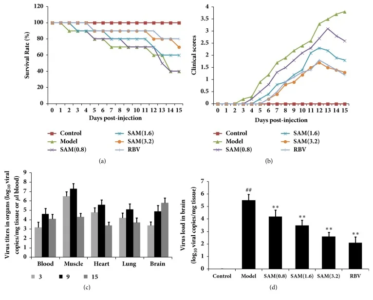
SAM reduced clinical scores, delayed disease development, and decreased levels of LDH, CK-MB, caspase 3, Bax, ER stress, and inflammatory reaction induced by CA16 infection.
SAM may attenuate CA16 brain injury in neonatal models; leaves open clinical translation for HFMD.
Original Research
FEB 2018ChenJournal of Neuroinflammation
IRE1α inhibition decreased TXNIP/NLRP3 inflammasome activation through miR-17-5p after neonatal hypoxic–ischemic brain injury in rats
BACKGROUND: The endoplasmic reticulum (ER) is responsible for the control of correct protein folding and protein function which is crucial for cell survival. However, under pathological conditions, such as hypoxia-ischemia (HI), there is an accumulation of unfolded proteins thereby triggering the unfolded protein response (UPR) and causing ER stress which is associated with activation of several stress sensor signaling pathways, one of them being the inositol requiring enzyme-1 alpha (IRE1α) signaling pathway. The UPR is regarded as a potential contributor to neuronal cell death and inflammation after HI. In the present study, we sought to investigate whether microRNA-17 (miR-17), a potential IRE1α ribonuclease (RNase) substrate, arbitrates downregulation of thioredoxin-interacting protein (TXNIP) and consequent NLRP3 inflammasome activation in the immature brain after HI injury and whether inhibition of IRE1α may attenuate inflammation via miR-17/TXNIP regulation. METHODS: ). STF-083010, an IRE1α RNase inhibitor, was intranasally delivered at 1 h post-HI or followed by an additional one administration per day for 2 days. MiR-17-5p mimic or anti-miR-17-5p inhibitor was injected intracerebroventricularly at 48 h before HI. Infarct volume and body weight were used to evaluate the short-term effects while brain weight, gross and microscopic brain tissue morphologies, and neurobehavioral tests were conducted for the long-term evaluation. Western blots, immunofluorescence staining, reverse transcription quantitative real-time polymerase chain reaction (RT-qPCR), and co-immunoprecipitation (Co-IP) were used for mechanism studies. RESULTS: Endogenous phosphorylated IRE1α expression was significantly increased after HI. Intranasal administration of STF-083010 alleviated brain injury and improved neurological behavior. MiR-17-5p expression was reduced after HI, and this decrease was attenuated by STF-083010 treatment. MiR-17-5p mimic administration ameliorated TXNIP expression, NLRP3 inflammasome activation, caspase-1 cleavage, and IL-1β production, as well as brain infarct volume. Conversely, anti-miR-17-5p inhibitor reversed IRE1α inhibition-induced decrease in TXNIP expression and inflammasome activation, as well as exacerbated brain injury after HI. CONCLUSIONS: IRE1a-induced UPR pathway may contribute to inflammatory activation and brain injury following neonatal HI. IRE1a activation, through decay of miR-17-5p, elevated TXNIP expression to activate NLRP3 inflammasome and aggravated brain damage.
Original Research
FEB 2017ChenMolecular Medicine Reports
99Tc-MDP-induced human osteoblast proliferation, differentiation and expression of osteoprotegerin
The aim of the present study was to examine the influence of technetium methylenediphosphonate (99Tc-MDP) on the proliferation and differentiation of human osteoblasts. Human iliac cancellous bone was isolated and cultured with either 99Tc‑MDP, β fibroblast growth factor (as a positive control) or medium only (as a negative control). Proliferation was assessed by direct cell counting, CCK‑8 assay and bromodeoxyuridine staining. The cell cycle and rate of apoptosis was assessed by propidium iodide staining and flow cytometry. Alkaline phosphatase (ALP) activity was assessed by the p‑nitrophenyl phosphate method and mineralized nodules were stained with Alizarin Red. Expression of osteocalcin (OCN) and bone morphogenetic protein‑2 (BMP‑2) was assessed by reverse transcription‑quantitative polymerase chain reaction (RT‑qPCR), and expression levels of osteoprotegerin (OPG) and receptor activator of NF‑κB ligand (RANKL) were assessed by RT‑qPCR and ELISA. Isolated human osteoblasts stained positively for ALP and developed mineralized nodules. Treatment with 10‑5‑10‑10 M 99Tc‑MDP enhanced proliferation and 48 h incubation with 10‑8 M 99Tc‑MDP increased the proportion of cells in S‑phase, decreased the proportion in G0/G1 phase, and increased the cell proliferation index. The rate of apoptosis also increased, but the increase was not significant. Cells incubated with 10‑6‑10‑9 M 99Tc‑MDP for 3‑9 days exhibited increased ALP activity and mineralized nodule development. 10‑8 M 99Tc‑MDP increased BMP‑2 and OPG expression levels and OPG secretion, but OCN mRNA expression levels and RANKL secretion were not significantly altered at 72 h. 99Tc‑MDP treatment induced osteoblast proliferation and differentiation without affecting apoptosis. These findings provide proof of concept for the future use of 99Tc‑MDP in the treatment of bone‑destructive diseases.
Original Research
JUL 2016GongMolecular Medicine Reports
Silencing of osterix expression by siRNA inhibits aldosterone-induced calcification of vascular smooth muscle cells in mice
The process of vascular calcification shares numerous similarities with that of skeletal mineralization and involves the deposition of hydroxyapatite crystals in arteries and cardiac valves. However, the underlying cellular mechanism remains to be fully elucidated. Microarray analysis in the present study demonstrated that greater than 2,000 genes were upregulated during the calcification of murine vascular smooth muscle cells (VSMCs), of which osterix (OSX) and integrin‑binding sialoprotein (IBSP) were the most significantly differentially expressed genes. Following the validation of increased OSX and IBSP expression by reverse transcription‑quantitative polymerase chain reaction in calcifying murine VSMCs induced by aldosterone. Subsequent to transfection with siRNA‑OSX, results indicated that OSX may inhibit calcification of VSMCs via IBSP. It was suggested that the increased OSX expression in calcifying VSMCs may reflect the well‑established prenatal role of OSX. A full understanding of the importance of OSX in this pathological process would improve understanding of the pathogenesis of vascular calcification.
Original Research
JAN 2016GuoStroke
Lipoxin A4 Reduces Inflammation Through Formyl Peptide Receptor 2/p38 MAPK Signaling Pathway in Subarachnoid Hemorrhage Rats
BACKGROUND AND PURPOSE: Lipoxin A4 (LXA4) has been reported to reduce inflammation in several neurological injury models. We studied the effects of LXA4 on neuroinflammation after subarachnoid hemorrhage (SAH) in a rat model. METHODS: Two hundred and thirty-eight Sprague-Dawley male rats, weight 280-320 g, were used. Exogenous LXA4 (0.3 and 1.0 nmol) were injected intracerebroventricularly at 1.5 hours after SAH. Neurological scores, brain water content, and blood-brain barrier were evaluated at 24 hours after SAH; Morris water maze and T-maze tests were examined at 21 days after SAH. The expression of endogenous LXA4 and its receptor formyl peptide receptor 2 (FPR2), as well as p38, interleukin-1β, and interleukin-6 were studied either by ELISA or by Western blots. Neutrophil infiltration was observed by myeloperoxidase staining. FPR2 siRNA was used to knock down LXA4 receptor. RESULTS: The expression of endogenous LXA4 decreased, and the expression of FPR2 increased after SAH. Exogenous LXA4 decreased brain water content, reduced Evans blue extravasation, and improved neurological functions and improved the learning and memory ability after SAH. LXA4 reduced neutrophil infiltration and phosphorylation of p38, interleukin-1β, and interleukin-6. These effects of LXA4 were abolished by FPR2 siRNA. CONCLUSIONS: Exogenous LXA4 inhibited inflammation by activating FPR2 and inhibiting p38 after SAH. LXA4 may serve as an alternative treatment to relieve early brain injury after SAH.
Original Research
DEC 2015TengStroke
Peroxisome Proliferator–Activated Receptor β/δ Alleviates Early Brain Injury After Subarachnoid Hemorrhage in Rats
BACKGROUND AND PURPOSE: Early brain injury is proposed to be the primary cause of the poor outcome after subarachnoid hemorrhage (SAH), which is closely related to the neural apoptosis. To date, the relationship between peroxisome proliferator-activated receptor β/δ (PPARβ/δ) and nuclear factor-κB/matrix metalloproteinase-9 (NF-κB/MMP-9) pathway, both of which are closely related to apoptotic effects, has been poorly studied in SAH. The present study was undertaken to evaluate the effects of PPARβ/δ on early brain injury and NF-κB/MMP-9 pathway after SAH in rats. METHODS: SAH model was established by injecting nonheparinized autologous arterial blood into the prechiasmatic cistern in male Sprague-Dawley rats. Adenoviruses or small interfering RNAs were injected into the right lateral cerebral ventricle to, respectively, up- or downregulate PPARβ/δ expression before SAH. All animals were assessed with a neurological score and then killed at 24 hours after SAH surgery. The indexes of brain water content, blood-brain barrier permeability, and apoptosis were used to detect brain injury. The expression of PPARβ/δ, NF-κB, and MMP-9 were measured by immunohistochemistry, gelatin zymography, and Western Blot methods, respectively. In addition, GW0742, a specific agonist of PPARβ/δ, was used to treat SAH in rats, the effects of which were evaluated by neurological scoring and Evans blue extravasation. RESULTS: Overexpression of PPARβ/δ by adenoviruses treatment significantly ameliorated brain injury with improvement in neurological deficits, brain edema, blood-brain barrier impairment, and neural cell apoptosis at 24 hours after SAH in rats, whereas downregulation of PPARβ/δ by small interfering RNAs administration resulted in the reverse effects of the above. The expression levels of NF-κB and MMP-9 were markedly downregulated when PPARβ/δ increased after PPARβ/δ adenovirus transfection and upregulated when PPARβ/δ decreased by PPARβ/δ small interfering RNAs treatment. Moreover, GW0742 improved neurological deficits and reduced Evans blue extravasation at 24 hours after SAH. CONCLUSIONS: PPARβ/δ's overexpression may attenuate early brain injury after rats' SAH administration, which reduces neural apoptosis possibly through blocking NF-κB/MMP-9 pathway.
Original Research
MAY 2015LiCritical Care Medicine
Progranulin Reduced Neuronal Cell Death by Activation of Sortilin 1 Signaling Pathways After Subarachnoid Hemorrhage in Rats
Objectives:Progranulin has been reported to have neuroprotective actions in cultured neurons. This study investigated the effect of recombinant rat progranulin on early brain injury after subarachnoid hemorrhage. Design:Controlled in vivo laboratory study. Setting:Animal research laboratory. Subjects:Two hundred thirty adult male Sprague-Dawley rats weighing 280–320 g. Interventions:Subarachnoid hemorrhage was induced in rats by endovascular perforation. Rat recombinant progranulin (1 and 3 ng) was administrated intracerebroventricularly at 1.5 hours after subarachnoid hemorrhage. Progranulin small interfering RNA was administrated by intracerebroventricularly at 1 day before subarachnoid hemorrhage induction. Subarachnoid hemorrhage grade, neurologic score, and brain water content were measured at 24 and 72 hours after subarachnoid hemorrhage. Neural apoptosis was evaluated by double immunofluorescence staining using terminal deoxynucleotidyl transferase-mediated uridine 5′-triphosphate-biotin nick-end labeling and neuronal nuclei. For mechanistic study, the expression of progranulin, phosphorylated Akt, Akt, p-Erk, Erk, Bcl-2, and cleaved caspase-3 were analyzed by Western blot at 24 hours after subarachnoid hemorrhage. siRNA for sortilin 1 (a progranulin receptor) was used to intervene the downstream pathway. Measurements and Main Results:The expression of progranulin decreased and reached the lowest point at 24 hours after subarachnoid hemorrhage. Administration of rat recombinant progranulin decreased brain water content and improved neurologic functions at both 24 and 72 hours after subarachnoid hemorrhage, while knockdown of endogenous progranulin aggravated neurologic deficits after subarachnoid hemorrhage. Rat recombinant progranulin treatment reduced neuronal apoptosis, while progranulin deficiency promoted neuronal apoptosis at 24 hours after subarachnoid hemorrhage. Rat recombinant progranulin promoted Akt activation, increased Bcl-2 level, but reduced caspase-3 level. Knockdown of progranulin binding factor sortilin 1 abolished the beneficial effects of rat recombinant progranulin at 24 hours after subarachnoid hemorrhage. Conclusion:Rat recombinant progranulin alleviated neuronal death via sortilin 1-mediated and Akt-related antiapoptosis pathway. Rat recombinant progranulin may have potentials to ameliorate early brain injury for subarachnoid hemorrhage patients.
Original Research
MAY 2015TianAndrologia
Effect of androgen deprivation on the expression of aquaporins in rat prostate and seminal vesicles
The aim of this study was to investigate the level of secretions of prostate and seminal vesicles and its association with the expression of AQP0, 1, 4, 5, 6 and 8 in castrated rats. Eight-week-old male Sprague-Dawley (SD) rats (n = 18) were randomly divided into control group, castrated rats group and castrated followed testosterone replacement group. Four weeks after surgery, the secretions and expression of AQP0, 1, 4, 5, 6 and 8 of prostate and seminal vesicles were determined. Serum testosterone was significantly lower in castrated groups than in control and testosterone replacement groups (P < 0.05). The level of prostate and seminal vesicle secretions and the expressions of AQP0, 1, 4, 5, 6 and 8 in prostate and seminal vesicles were significantly lower in castrated group than in control and castrated followed testosterone replacement groups (P < 0.05). The decreased prostatic and seminal vesicle secretions in castrated rats may be related to the decrease in AQP0, 1, 4, 5, 6 and 8 in prostatic tissue and seminal vesicles.