IJ
Ian Judson
Cardio-Oncology
Institute of Cancer Research
About
Ian Judson works in Cardio-Oncology affiliated with Institute of Cancer Research.
Ian Judson is Emeritus Professor at the Institute of Cancer Research in London. He previously served as Professor of Cancer Pharmacology at the Institute of Cancer Research and as a Consultant Medical Oncologist at the Royal Marsden Hospital. His research includes sarcoma oncology with a focus on cardiac tumors and thrombi.
Research focus
Cardiac SarcomasAngiosarcomasChemotherapy Cardiac SafetyVascular TumorsCardiac Tumor Treatment
Impact
591 pubs · 46,454 cites · h-index 100
Training
MD
University of Cambridge
1988
MA
University of Cambridge
Affiliations
Work Experience
Emeritus Professor
Institute of Cancer Research · 2016 - Present
Professor of Cancer Pharmacology
Institute of Cancer Research · 1989 - 2016
Consultant Medical Oncologist
Royal Marsden Hospital · 1989 - 2016
Publications
Risk Assessment
JUN 2024JudsonBritish Journal of Cancer
Gastrointestinal stromal tumour (GIST): British Sarcoma Group clinical practice guidelines
Guidelines represent a consensus view of current best clinical practice. Where appropriate, key recommendations are given and the levels of evidence and strength of recommendation gradings are those used by the European Society for Medical Oncology (ESMO).
Clinical Trial
MAY 2024HayesBritish Journal of Cancer
UK guidelines for the management of soft tissue sarcomas
Soft tissue sarcomas (STS) are rare tumours arising in mesenchymal tissues and can occur almost anywhere in the body. Their rarity, and the heterogeneity of subtype and location, means that developing evidence-based guidelines is complicated by the limitations of the data available. This makes it more important that STS are managed by expert multidisciplinary teams, to ensure consistent and optimal treatment, recruitment to clinical trials, and the ongoing accumulation of further data and knowledge. The development of appropriate guidance, by an experienced panel referring to the evidence available, is therefore a useful foundation on which to build progress in the field. These guidelines are an update of the previous versions published in 2010 and 2016 [1, 2]. The original guidelines were drawn up by a panel of UK sarcoma specialists convened under the auspices of the British Sarcoma Group (BSG) and were intended to provide a framework for the multidisciplinary care of patients with soft tissue sarcomas. This iteration of the guidance, as well as updating the general multidisciplinary management of soft tissue sarcoma, includes specific sections relating to the management of sarcomas at defined anatomical sites: gynaecological sarcomas, retroperitoneal sarcomas, breast sarcomas, and skin sarcomas. These are generally managed collaboratively by site specific multidisciplinary teams linked to the regional sarcoma specialist team, as stipulated in the recently published sarcoma service specification [3]. In the UK, any patient with a suspected soft tissue sarcoma should be referred to a specialist regional soft tissues sarcoma service, to be managed by a specialist sarcoma multidisciplinary team. Once the diagnosis has been confirmed using appropriate imaging and a tissue biopsy, the main modality of management is usually surgical excision performed by a specialist surgeon, combined with pre- or post-operative radiotherapy for tumours at higher risk for local recurrence. Systemic anti-cancer therapy (SACT) may be utilised in cases where the histological subtype is considered more sensitive to systemic treatment. Regular follow-up is recommended to assess local control, development of metastatic disease, and any late effects of treatment.
Cohort Study
SEP 2023CalvertJournal of Clinical Oncology
Carboplatin dosage: prospective evaluation of a simple formula based on renal function.
A dosage formula has been derived from a retrospective analysis of carboplatin pharmacokinetics in 18 patients with pretreatment glomerular filtration rates (GFR) in the range of 33 to 136 mL/min. Carboplatin plasma clearance was linearly related to GFR (r = 0.85, P less than .00001) and rearrangements of the equation describing the correlation gave the dosage formula dose (mg) = target area under the free carboplatin plasma concentration versus time curve (AUC) x (1.2 x GFR + 20). In a prospective clinical and pharmacokinetic study the formula was used to determine the dose required to treat 31 patients (GFR range, 33 to 135 mL/min) with 40 courses of carboplatin. The target AUC was escalated from 3 to 8 mg carboplatin/mL/min. Over this AUC range the formula accurately predicted the observed AUC (observed/predicted ratio 1.24 +/- 0.11, r = 0.886) and using these additional data, the formula was refined. Dose (mg) = target AUC x (GFR + 25) is now the recommended formula. AUC values of 4 to 6 and 6 to 8 mg/mL. min gave rise to manageable hematological toxicity in previously treated and untreated patients, respectively, and hence target AUC values of 5 and 7 mg/mL min are recommended for single-agent carboplatin in these patient groups. Pharmacokinetic modeling demonstrated that the formula was reasonably accurate regardless of whether a one- or two-compartment model most accurately described carboplatin pharmacokinetics, assuming that body size did not influence nonrenal clearance. The validity of this assumption was demonstrated in 13 patients where no correlation between surface area and nonrenal clearance was found (r = .31, P = .30). Therefore, the formula provides a simple and consistent method of determining carboplatin dose in adults. Since the measure of carboplatin exposure in the formula is AUC, and not toxicity, it will not be influenced by previous or concurrent myelosuppressive therapy or supportive measures. The formula is therefore applicable to combination and high-dose studies as well as conventional single-agent therapy, although the target AUC for carboplatin will need to be redefined for combination chemotherapy.
Human Clinical Trial
SEP 2023WollBritish Journal of Cancer
Axitinib in patients with advanced/metastatic soft tissue sarcoma (Axi-STS): an open-label, multicentre, phase II trial in four histological strata
BACKGROUND: Axitinib is an oral vascular endothelial growth factor receptor inhibitor with anti-tumour activity in renal, thyroid, and pancreatic cancer. METHODS: Axi-STS was a pathologically-stratified, non-randomised, open-label, multi-centre, phase II trial of continuous axitinib treatment in patients ≥16 years, performance status ≤2, with pathologically-confirmed advanced/metastatic soft tissue sarcoma (STS). Patients were recruited within four tumour strata, each analysed separately: angiosarcoma, leiomyosarcoma, synovial sarcoma, or other eligible STSs. The primary outcome was progression-free survival at 12 weeks (PFS12). A Simon's two-stage design with activity defined as PFS12 rate of 40% determined a sample size of 33 patients per strata. RESULTS: Between 31-August-2010 and 29-January-2016, 145 patients were recruited: 38 angiosarcoma, 37 leiomyosarcoma, 36 synovial sarcoma, and 34 other subtypes. PFS12 rate for each stratum analysed was 42% (95% lower confidence interval (LCI); 29), 45% (95% LCI; 32), 57% (95% LCI; 42), and 33% (95% LCI; 21), respectively. There were 74 serious adverse events including two treatment-related deaths of pulmonary haemorrhage and gastrointestinal bleeding. Fatigue and hypertension were the most common grade 3 adverse events. CONCLUSIONS: Axitinib showed clinical activity in all STS strata investigated. The adverse event profile was acceptable, supporting further investigation in phase III trials. CLINICAL TRIAL REGISTRATION: ISRCTN 60791336.
Original Research
JUN 2023BurnsNature Communications
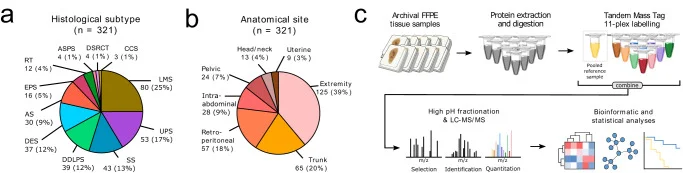
High expression of a vesicle transport protein signature (SPM10) was an independent positive prognostic factor for metastasis-free survival in soft tissue sarcomas (HR 0.479).
Provides a hypothesis-generating molecular taxonomy for rare sarcomas; leaves open whether these.
Early-Phase Clinical Trial
MAR 2023BairdErasmus MC
Data from Phase I safety, pharmacokinetic, and pharmacogenomic trial of ES-285, a novel marine cytotoxic agent, administered to adult patients with advanced solid tumors
Abstract A dose-escalation, phase I study evaluated the safety, pharmacokinetics, pharmacogenomics, and efficacy of ES-285, a novel agent isolated from a marine mollusc, in adult cancer patients. Patients received a 24-hour i.v. infusion of ES-285 once every 3 weeks until disease progression or unacceptable toxicity. The starting dose was 4 mg/m². Dose escalation in cohorts of at least three patients proceeded according to the worst toxicity observed in the previous cohort. Twenty-eight patients were treated with 72 courses of ES-285 across eight dose levels. No dose-limiting toxicities were seen between 4 and 128 mg/m². Two of four patients treated at 256 mg/m² had dose-limiting reversible grade 3 transaminitis; one patient at 256 mg/m² also had transient grade 3 central neurotoxicity. One of three patients subsequently treated at 200 mg/m² died following drug-related central neurotoxicity. Other toxicities included phlebitis, nausea, fatigue, and fever. Pharmacokinetic studies indicated dose proportionality with high volume of distribution (median Vss at 256 mg/m² was 2,389 liters; range, 1,615–4,051 liters) and long elimination half life (median t1/2 at 256 mg/m² was 28 h; range, 21–32 h). The three patients with dose-limiting toxicity had the highest drug exposure. Pharmacogenomic studies of paired surrogate tissue samples identified changes in gene expression following treatment that correlated with increasing dose. Disease stabilization for 6 to 18 weeks was recorded in nine patients. Using this schedule, 128 mg/m² was considered safe and feasible. At this dose, pharmacologically relevant concentrations of the drug were safely achieved with pharmacogenomic studies indicating changes in the expression of genes of potential mechanistic relevance. [Mol Cancer Ther 2009;8(6):1430–7]
Original Research
JAN 2023BallingerScience
Heritable defects in telomere and mitotic function selectively predispose to sarcomas
Cancer genetics has to date focused on epithelial malignancies, identifying multiple histotype-specific pathways underlying cancer susceptibility. Sarcomas are rare malignancies predominantly derived from embryonic mesoderm. To identify pathways specific to mesenchymal cancers, we performed whole-genome germline sequencing on 1644 sporadic cases and 3205 matched healthy elderly controls. Using an extreme phenotype design, a combined rare-variant burden and ontologic analysis identified two sarcoma-specific pathways involved in mitotic and telomere functions. Variants in centrosome genes are linked to malignant peripheral nerve sheath and gastrointestinal stromal tumors, whereas heritable defects in the shelterin complex link susceptibility to sarcoma, melanoma, and thyroid cancers. These studies indicate a specific role for heritable defects in mitotic and telomere biology in risk of sarcomas.
Guideline
NOV 2022RothermundtEuropean Journal of Cancer
Controversies in the management of patients with soft tissue sarcoma: Recommendations of the Conference on State of Science in Sarcoma 2022
BACKGROUND: Owing to the rarity and heterogeneity in biology and presentation, there are multiple areas in the diagnosis, treatment and follow-up of soft tissue sarcoma (STS), with no, low-level or conflicting evidence. METHODS: During the first Consensus Conference on the State of Science in Sarcoma (CSSS), we used a modified Delphi process to identify areas of controversy in the field of sarcoma, to name topics with limited evidence-based data in which a scientific and knowledge gap may remain and a consensus statement will help to guide patient management. We determined scientific questions which need to be addressed in the future in order to generate evidence and to inform physicians and caregivers in daily clinical practice in order to improve the outcomes of patients with sarcoma. We conducted a vote on STS key questions and controversies prior to the CSSS meeting, which took place in May 2022. RESULTS: Sixty-two European sarcoma experts participated in the survey. Sixteen strong consensus (≥95%) items were identified by the experts, as well as 30 items with a ≥75% consensus on diagnostic and therapeutic questions. Ultimately, many controversy topics remained without consensus. CONCLUSIONS: In this manuscript, we summarise the voting results and the discussion during the CSSS meeting. Future scientific questions, priorities for clinical trials, registries, quality assurance, and action by stakeholders are proposed. Platforms and partnerships can support innovative approaches to improve management and clinical research in STS.
Human Clinical Trial
SEP 2022KantidakisEuropean Journal of Cancer
New benchmarks to design clinical trials with advanced or metastatic liposarcoma or synovial sarcoma patients: An EORTC – Soft Tissue and Bone Sarcoma Group (STBSG) meta-analysis based on a literature review for soft-tissue sarcomas
BACKGROUND: Recently, we performed a meta-analysis based on a literature review for STS trials (published 2003-2018, ≥10 adult patients) to update long-standing reference values for leiomyosarcomas. This work is extended for liposarcomas (LPS) and synovial sarcomas (SS). MATERIALS AND METHODS: Study endpoints were progression-free survival rates (PFSRs) at 3 and 6 months. Trial-specific estimates were pooled per treatment line (first-line or pre-treated) with random effects meta-analyses. The choice of the therapeutic benefit to target in future trials was guided by the European Society for Medical Oncology Magnitude of Clinical Benefit Scale (ESMO-MCBS). RESULTS: Information was acquired for 1030 LPS patients (25 trials; 7 first-line, 17 pre-treated, 1 both) and 348 SS patients (13 trials; 3 first-line, 10 pre-treated). For LPS, the overall pooled first-line PFSRs were 69% (95%-CI 60-77%) and 56% (95%-CI 45-67%) at 3 and 6 months, respectively. These rates were 49% (95%-CI 40-57%)/28% (95%-CI 22-34%) for >1 lines. For SS, first-line PFSRs were 74% (95%-CI 58-86%)/56% (95%-CI 31-78%) at 3 and 6 months, and pre-treated rates were 45% (95%-CI 34-57%)/25% (95%-CI 16-36%). Following ESMO-MCBS guidelines, the minimum values to target are 79% and 69% for first-line LPS (82% and 69% for SS) at 3 and 6 months. For pre-treated LPS, recommended PFSRs at 3 and 6 months suggesting drug activity are 63% and 44% (60% and 41% for SS). CONCLUSIONS: New benchmarks are proposed for advanced/metastatic LPS or SS to design future histology-specific phase II trials. More data are needed to provide definitive thresholds for the different LPS subtypes.
Original Research
JUN 2022KrásnýCancers
Proteomic Profiling Identifies Co-Regulated Expression of Splicing Factors as a Characteristic Feature of Intravenous Leiomyomatosis
Intravenous leiomyomatosis (IVLM) is a rare benign smooth muscle tumour that is characterised by intravenous growth in the uterine and pelvic veins. Previous DNA copy number and transcriptomic studies have shown that IVLM harbors unique genomic and transcriptomic alterations when compared to uterine leiomyoma (uLM), which may account for their distinct clinical behaviour. Here we undertake the first comparative proteomic analysis of IVLM and other smooth muscle tumours (comprising uLM, soft tissue leiomyoma and benign metastasizing leiomyoma) utilising data-independent acquisition mass spectrometry. We show that, at the protein level, IVLM is defined by the unique co-regulated expression of splicing factors. In particular, IVLM is enriched in two clusters composed of co-regulated proteins from the hnRNP, LSm, SR and Sm classes of the spliceosome complex. One of these clusters (Cluster 3) is associated with key biological processes including nascent protein translocation and cell signalling by small GTPases. Taken together, our study provides evidence of co-regulated expression of splicing factors in IVLM compared to other smooth muscle tumours, which suggests a possible role for alternative splicing in the pathogenesis of IVLM.
cohort
MAY 2022StacchiottiCancer

Ifosfamide-containing regimens showed higher activity compared to anthracycline alone, although it was not statistically significant.
Does not support changing first-line anthracycline use; leaves open whether ifosfamide addition improves activity in advanced liposarcoma.
Original Research
APR 2022KantidakisSarcoma
Prognostic Significance of Bone Metastasis in Soft Tissue Sarcoma Patients Receiving Palliative Systemic Therapy: An Explorative, Retrospective Pooled Analysis of the EORTC-Soft Tissue and Bone Sarcoma Group (STBSG) Database
Background: Soft-tissue sarcomas (STS) constitute a rare group of heterogeneous mesenchymal tumours containing more than 100 histologic subtypes. Here, we investigate whether, and if so, to what extent, skeletal metastases affect the outcome of patients with advanced or metastatic disease. Materials and Methods: Selected patients participated in five clinical trials of EORTC-STBSG. Individuals were included if they started treatment with an active drug and had advanced/metastatic STS. The endpoints of interest were overall survival (OS) and progression-free survival (PFS). Univariate and multivariate pooled analyses (after correcting for 12 covariates) were employed with Kaplan-Meier and Cox regression to model the impact of bone metastasis at presentation per treatment line stratified by study. For the subset of patients with bone metastasis, the impact of another metastatic organ site was explored with multivariate Cox regression models. Results: 565 out of 1034 (54.6%) patients received first-line systemic treatment for locally advanced or metastatic disease. Bone metastases were present in 140 patients (77 first-line, 63 second-line or higher). The unadjusted difference in OS/PFS with or without bone metastasis was statistically significant only for first-line patients. For OS, the adjusted hazard ratios for bone metastasis presence were 1.33 (95%-CI: 0.99-1.78) and 1.11 (95%-CI: 0.81-1.52) for first-line/second-line or higher treated patients, respectively. Likewise, the adjusted hazard ratios for PFS were 1.31 (95%-CI: 1.00-1.73) and 1.07 (95%-CI: 0.80-1.43). Effects were not statistically significant, despite a trend in first-line patients for both endpoints. Subgroup analyses indicated bone and lymph node metastasis as the most detrimental combination for OS and bone and lung metastasis for PFS. Conclusions: Adult STS patients receiving palliative systemic therapy with bone metastasis carried an overall worse prognosis than STS patients without bone metastases. Skeletal metastasis was detrimental for both OS and PFS, independent of the treatment line. Findings may have implications for the management of these patients.
Original Research
MAR 2022KrásnýInstitute of Cancer Research
Proteomic profiling identifies co-regulated expression of splicing factors as a characteristic feature of intravenous leiomyomatosis
Intravenous leiomyomatosis (IVLM) is a rare benign smooth muscle tumour that is characterised by intravenous growth in the uterine and pelvic veins. Previous DNA copy number and transcriptomic studies have shown that IVLM harbours unique genomic and transcriptomic alterations when compared to uterine leiomyoma (uLM), which may account for their distinct clinical behaviour. Here we undertake the first comparative proteomic analysis of IVLM and other smooth muscle tumours (comprising uLM, soft tissue leiomyoma and benign metastasising leiomyoma) utilising data-independent acquisition mass spectrometry. We show that, at the protein level, IVLM is defined by the unique co-regulated expression of splicing factors. In particular, IVLM is enriched in two clusters composed of co-regulated proteins from the hnRNP, LSm, SR and Sm classes of the spliceosome complex. One of these clusters (Cluster 3) is associated with key biological processes including nascent protein translocation and cell signalling by small GTPases. Taken together, our study provides evidence of co-regulated expression of splicing factors in IVLM compared to other smooth muscle tumours which suggests a possible role for alternative splicing in the pathogenesis of IVLM.
Prognostic Study
SEP 2021WallsAnticancer Research
Treatments and Outcomes in Oligometastatic Soft Tissue Soft Sarcoma – A Single Centre Retrospective Analysis
BACKGROUND/AIM: Distinguishing true oligometastatic disease from early polymetastatic disease is vital in patients with soft tissue sarcoma as contemporary treatment strategies differ significantly. Clinical factors such as tumour biology, organ involved, number of lesions, and patient fitness influence clinical decisions. PATIENTS AND METHODS: A retrospective search of a prospective database identified patients with new distant relapse, treated between 2009 and 2012. RESULTS: A total of 223 patients were included, and oligometastases were diagnosed in 81 (36%) patients, which were pulmonary in just over half of cases. These were treated with local therapy in 66 of 89 cases, and 7 patients received subsequent treatment for additional oligometastases. Metastasectomy was the most common treatment modality. A total of 16/66 patients (24%) underwent active surveillance for >6 months prior to local therapy. CONCLUSION: Patients with oligometastatic disease can experience durable disease control with timely multimodality treatment approaches for evolving metastatic disease, where disease biology allows.
Human Clinical Trial
JUL 2021KantidakisEuropean Journal of Cancer
Efficacy thresholds for clinical trials with advanced or metastatic leiomyosarcoma patients: A European Organisation for Research and Treatment of Cancer Soft Tissue and Bone Sarcoma Group meta-analysis based on a literature review for soft-tissue sarcomas
BACKGROUND: In 2002, the European Organisation for Research and Treatment of Cancer Soft Tissue and Bone Sarcoma Group reported well-established values for conducting phase II trials for soft-tissue sarcomas. An update is provided for leiomyosarcoma (LMS). MATERIALS AND METHODS: Clinical trials with advanced or metastatic LMS were identified via literature review in PubMed (published 2003-2018, ≥10 adult LMS patients). End-points were 3- and 6-month progression-free survival rates (PFSR-3m and PFSR-6m). When estimates could not be derived from publications, data requests were sent out. Treatments were classified as recommended (R-T) or non-recommended (NR-T) according to the ESMO 2018 guidelines. A random effects meta-analysis was used to pool trial-specific estimates for first-line (1L) or pre-treated (2L+) patients separately. The ESMO Magnitude of Clinical Benefit Scale was used to guide the treatment effect to target in future trials. RESULTS: From 47 studies identified, we obtained information on 7 1L and 16 2L+ trials for 1500 LMS patients. Overall, in 1L, PFSR-3m and PFSR-6m were 74% (95% confidence interval [CI] 64-82%) and 58% (95% CI 50-66%), respectively. For 2L+, PFSR-3m was 48% (95% CI 41-54%), and PFSR-6m was 28% (95% CI 22-34%). No difference was observed between R-T and NR-T for first or later lines. Under the alternative that the true benefit amounts to a hazard ratio of 0.65, a PFSR-6m ≥70% can be considered to suggest drug activity in 1L. For 2L+, a PFSR-3m ≥62% or PFSR-6m ≥44% would suggest drug activity. Specific results are also provided for uterine LMS. CONCLUSIONS: This work provides a new benchmark for designing phase II studies for advanced or metastatic LMS.
Original Research
APR 2021MilighettiJournal of Proteomics
Proteomic profiling of soft tissue sarcomas with SWATH mass spectrometry
Soft tissue sarcomas (STS) are a group of rare and heterogeneous cancers. While large-scale genomic and epigenomic profiling of STS have been undertaken, proteomic analysis has thus far been limited. Here we utilise sequential window acquisition of all theoretical fragment ion spectra mass spectrometry (SWATH-MS) for proteomic profiling of formalin fixed paraffin embedded (FFPE) specimens from a cohort of STS patients (n = 36) across four histological subtypes (leiomyosarcoma, synovial sarcoma, undifferentiated pleomorphic sarcoma and dedifferentiated liposarcoma). We quantified 2951 proteins across all cases and show that there is a significant enrichment of gene sets associated with smooth muscle contraction in leiomyosarcoma, RNA splicing regulation in synovial sarcoma and leukocyte activation in undifferentiated pleomorphic sarcoma. We further identified a subgroup of STS cases that have a distinct expression profile in a panel of proteins, with worse survival outcomes when compared to the rest of the cohort. Our study highlights the value of comprehensive proteomic characterisation as a means to identify histotype-specific STS profiles that describe key biological pathways of clinical and therapeutic relevance; as well as for discovering new prognostic biomarkers in this group of rare and difficult-to-treat diseases.
Case Report
DEC 2020JudsonGynecologic Oncology Reports
Vitamin D deficiency in the pathogenesis of leiomyoma and intravascular leiomyomatosis: A case report and review of the literature
This is a case of recurrent intravascular leiomyomatosis in a pre-menopausal woman of African-Caribbean heritage. She presented in 2006 with multiple uterine leiomyomata, tumour invading the inferior vena cava (IVC) extending into the right atrium, and pulmonary metastases. Her initial presentation was treated surgically. On recurrence she was treated by oestrogen suppression using a combination of goserelin and letrozole, with a substantial response. She subsequently reported further regression of disease following exposure to strong sunlight enabling her to discontinue oestrogen suppression without relapse. The hypothesis is that the benefit was due to vitamin D. The role of hypovitaminosis D in the pathogenesis of uterine leiomyomata is discussed, including epidemiology data demonstrating a link between ethnicity and risk and the proven mechanisms by which vitamin D controls oestrogen and progesterone receptor expression and influences other signalling pathways involved in the pathogenesis of leiomyomas. Data indicating the intermediate malignancy nature of intravascular leiomyomatosis, are discussed. We are not aware of other reports indicating a link between intravascular leiomyomatosis and a lack of vitamin D.
Original Research
SEP 2020DemetriTyöväentutkimus Vuosikirja

Diagnosis and management of tropomyosin receptor kinase (TRK) fusion sarcomas: expert recommendations from the World Sarcoma Network
Sarcomas are a heterogeneous group of malignancies with mesenchymal lineage differentiation. The discovery of neurotrophic tyrosine receptor kinase (NTRK) gene fusions as tissue-agnostic oncogenic drivers has led to new personalized therapies for a subset of patients with sarcoma in the form of tropomyosin receptor kinase (TRK) inhibitors. NTRK gene rearrangements and fusion transcripts can be detected with different molecular pathology techniques, while TRK protein expression can be demonstrated with immunohistochemistry. The rarity and diagnostic complexity of NTRK gene fusions raise a number of questions and challenges for clinicians. To address these challenges, the World Sarcoma Network convened two meetings of expert adult oncologists and pathologists and subsequently developed this article to provide practical guidance on the management of patients with sarcoma harboring NTRK gene fusions. We propose a diagnostic strategy that considers disease stage and histologic and molecular subtypes to facilitate routine testing for TRK expression and subsequent testing for NTRK gene fusions.
Prognostic Study
MAY 2020YoungerJournal of Adolescent and Young Adult Oncology
Metastatic Soft Tissue Sarcomas in Adolescents and Young Adults: A Specialist Center Experience
Purpose: Soft tissue sarcomas (STS) account for 8% of all cancers in adolescents and young adults (AYAs). Metastatic STS contribute significantly to disease-related mortality in this age group; however, data are limited due to under-representation in clinical trials. Methods: AYAs aged 18–39 years, diagnosed with metastatic STS between 1990 and 2012, were identified from The Royal Marsden Hospital database. Outcomes of interest were clinical characteristics, treatment patterns, overall survival (OS), and prognostic factors. Results: Overall, 455 patients were included. Median age at diagnosis of metastatic STS was 33 years (interquartile range [IQR] 27–37 years). The most common histological subtypes were leiomyosarcoma ( n = 68, 15%), synovial sarcoma ( n = 68, 15%), Ewing sarcoma ( n = 44, 10%), and rhabdomyosarcoma ( n = 35, 8%). Treatments included systemic therapy ( n = 395, 87%; median 2 lines [IQR 1–3]; clinical trial n = 93, 22%), radiotherapy ( n = 297, 66%), and metastasectomy ( n = 191, 43%). Median duration between last chemotherapy regimen and death was 4.6 months (IQR 2–10). Median OS was 19.2 months (95% confidence interval [CI] 15.8–22.2); 5-year OS was 16%. Of common subtypes, patients with rhabdomyosarcoma had the worst OS (8.8 months; 95% CI 7.9–11.4). Adverse prognostic factors included male gender, synchronous metastases, bone or liver metastases, first-line polychemotherapy, and no metastasectomy. Conclusions: Outcomes were variable; patients with supposed chemosensitive subtypes had particularly poor survival. The diverse behavior of STS in AYAs highlights the need for subtype-specific clinical trials.
RCT
APR 2020GronchiJAMA Surgery

Quality of Surgery and Outcome in Localized Gastrointestinal Stromal Tumors Treated Within an International Intergroup Randomized Clinical Trial of Adjuvant Imatinib
Importance: The association between quality of surgery and overall survival in patients affected by localized gastrointestinal stromal tumors (GIST) is not completely understood. Objective: To assess the risk of death with and without imatinib according to microscopic margins status (R0/R1) using data from a randomized study on adjuvant imatinib. Design, Setting, and Participants: This is a post hoc observational study on patients included in the randomized, open-label, phase III trial, performed between December 2004 and October 2008. Median follow-up was 9.1 years (IQR, 8-10 years). The study was performed at 112 hospitals in 12 countries. Inclusion criteria were diagnosis of primary GIST, with intermediate or high risk of relapse; no evidence of residual disease after surgery; older than 18 years; and no prior malignancies or concurrent severe/uncontrolled medical conditions. Data were analyzed between July 17, 2017, and March 1, 2020. Interventions: Patients were randomized after surgery to either receive imatinib (400 mg/d) for 2 years or no adjuvant treatment. Randomization was stratified by center, risk category (high vs intermediate), tumor site (gastric vs other), and quality of surgery (R0 vs R1). Tumor rupture was included in the R1 category but also analyzed separately. Main Outcomes and Measures: Primary end point of this substudy was overall survival (OS), estimated using Kaplan-Meier method and compared between R0/R1 using Cox models adjusted for treatment and stratification factors. Results: A total of 908 patients were included; 51.4% were men (465) and 48.6% were women (440), and the median age was 59 years (range, 18-89 years). One hundred sixty-two (17.8%) had an R1 resection, and 97 of 162 (59.9%) had tumor rupture. There was a significant difference in OS for patients undergoing an R1 vs R0 resection, overall (hazard ratio [HR], 2.05; 95% CI, 1.45-2.89) and by treatment arm (HR, 2.65; 95% CI, 1.37-3.75 with adjuvant imatinib and HR, 1.86; 95% CI, 1.16-2.99 without adjuvant imatinib). When tumor rupture was excluded, this difference in OS between R1 and R0 resections disappeared (HR, 1.05; 95% CI, 0.54-2.01). Conclusions and Relevance: The difference in OS by quality of surgery with or without imatinib was associated with the presence of tumor rupture. When the latter was excluded, the presence of R1 margins was not associated with worse OS. Trial Registration: ClinicalTrials.gov Identifier: NCT00103168.