TD
T. Dylan Olver
Cardiovascular and exercise physiology
University of Saskatchewan
About
Research focus
Cardiovascular and exercise physiology
Impact
126 pubs · 1,700 cites · h-index 22
Affiliations
Publications
Retrospective Study
SEP 2025LuchkanychJournal of the American Heart Association
Effect of Phenylephrine on Cerebrovascular Regulation: A Translational Perspective
Retrospective analysis reveals phenylephrine affects cerebrovascular dynamics in humans and swine, indicating endothelial NO's role.
Original Research
JUN 2025DelgadoMicrocirculation
Modulatory Role of Nitric Oxide on the Vasomotor Actions of NPY in Porcine Cerebral Arteries
Neuropeptide Y (NPY) is a sympathetic co-transmitter that mediates vasoconstriction. However, there is evidence that it may also mediate dilation through a nitric oxide (NO)-dependent mechanism. OBJECTIVE: We used a swine model to examine how NPY influences cerebral vascular regulation and hypothesized that NPY would elicit both vasoconstrictor and vasodilatory effects, and that such effects would be modulated partially by NO signaling. Separately, Y1 receptor distribution (immunohistochemistry) and vasomotor responses to intra- and extraluminal NPY under control and NOS inhibition conditions were examined in isolated arteries. RESULTS: Intracarotid NPY infusions elicited transient dilation that was blocked by NOS inhibition. In isolated pial arteries, distinct populations of NPY-Y1 receptors were observed on both the vascular smooth muscle (VSM) and endothelium. Extraluminal application of NPY elicited vasoconstriction, while intraluminal delivery elicited vasodilation. NOS inhibition enhanced the magnitude of vasoconstriction in isolated pial arteries. Endothelial denudation, Y1 receptor antagonism, and NOS inhibition each blunted NPY-induced vasodilation. CONCLUSION: These data suggest both vasoconstrictor and vasodilatory effects of NPY are modulated partially by NO signaling.
case-control
JUN 2025KhanAJP Regulatory Integrative and Comparative Physiology
Acute isometric handgrip exercise failed to augment brachial artery flow-mediated dilation in young patients with congenital heart disease compared to healthy controls.
May indicate stress-induced endothelial dysfunction in congenital heart disease; leaves open utility of handgrip testing pending prospective validation.
cross-sectional
MAR 2025MorseJournal of Applied Physiology
Cardiac dysfunction markers including EF and ESVi are linked to brain atrophy and cognitive impairment.
Supports cardiac-brain link in HFrEF; hypothesis-generating, leaves open causality and practice implications.
case-control
MAR 2025JurrissenJournal of Applied Physiology
HFrEF patients display reduced resting cerebral perfusion but increased perfusion during PECO versus controls.
Altered cerebral perfusion responses in HFrEF are hypothesis-generating; leaves open whether metaboreflex modulation affects outcomes or warrants intervention.
Original Research
JUL 2024BoamahNeuroToxicology
Impaired memory in Sprague-Dawley rats exposed to complex groundwater mixtures of contaminants is associated with reduced cranial blood flow and hippocampal neurotoxicity
Exposure to industrial contaminants has been implicated in neurobehavioral toxicity in humans. To explore this potential risk, we investigated the neurotoxic effects of oral exposure to a complex groundwater mixture containing petroleum hydrocarbons, pesticides, heavy metals, and unknown parent and breakdown products using male and female Sprague Dawley rats. Rats were randomly divided into six groups and orally exposed daily via drinking water to: (i) tap water, (ii) 10 % v/v low impact groundwater, and (iii) 0.01 %, 0.1 %, 1 %, and 10 % high-impact groundwater for 60 days. Medium- and long-term memory (measured using the novel object recognition task) were impaired. However, no gross motor or coordination deficits were observed by the end of the study period (rotarod test). Doppler ultrasound of the middle cerebral and common carotid arteries was performed to examine the hemodynamic changes. The common carotid blood flow decreased in the groundwater-exposed rats compared to that in the control. However, no significant differences in cerebral blood velocity were observed between the exposed and control groups. A significant reduction in hippocampal serotonin levels was observed in groundwater-exposed rats relative to that in the control group. Collectively, these results indicate that impaired recognition memory in rats exposed to groundwater is accompanied by reduced cranial blood flow and hippocampal neurotoxicity, characterized by altered serotonergic signalling. The levels of detected contaminants known to cause neural or vascular damage were of magnitudes lower than the concentrations of contaminants found in the groundwater mixture, meaning the culprit chemical identity remains unknown. This study emphasizes the need to use whole mixture in exposures when dealing with complex contaminated sites rather than the use of individual compounds.
Original Research
MAY 2024BoyesAJP Heart and Circulatory Physiology

Elevated sympathetic-mediated vasoconstriction at rest but intact functional sympatholysis during exercise in heart failure with reduced ejection fraction
Review
MAY 2024BoyesPhysiology
Sex Differences in Left Ventricular Stroke Volume During Muscle Metaboreflex Activation
Introduction: Muscle metaboreflex activation (MMA) increases blood pressure, possibly by disproportionate increases in left ventricular (LV) stroke volume (SV) in males compared to females due to greater grip strength and metabolite production. The purpose of this study was to determine the effect of sex on SV during isolated MMA using cardiac MRI in young healthy adults. We hypothesized that SV would increase during MMA to a greater degree in males compared to females, and that this effect would be negated by accounting for grip strength. Methods: Ten males [23(2) yrs] and 10 females [26(5) yrs] underwent cardiac MRI during rest, 30% maximal voluntary contraction (MVC) isometric handgrip exercise (6 min), and isolation of MMA with brachial cuff circulatory occlusion (6 min). SV, end-systolic (ESV) and end-diastolic volumes (EDV) were measured by 2- and 4-chamber cine images and indexed (i) to body surface area to account for body size. Heart rate (HR, ECG) and mean arterial pressure (MAP, brachial cuff) were recorded. Cardiac output index (COi) and total peripheral resistance (TPR) were calculated. Analyses included ANOVA (sex × condition), ANCOVA [100% MVC (M=49(13) kg, F=26(7) kg) covariate], and linear regression. Results: SVi decreased from rest to MMA in males [67(8) to 61(5) mL/m 2 , p=0.0001] but not females [59(9) to 59(7) mL/m 2 , p=0.998; interaction p=0.002]. Notably, the reduction in SVi during MMA in males ( p=0.002) but not females ( p=0.957) remained with ANCOVA using MVC as the covariate (interaction p=0.021). ESVi increased during MMA in males [39(5) to 48(10) mL/m 2 , p<0.0001] but not females [32(5) to 36(6) mL/m 2 , p=0.092; interaction p=0.029]. MMA decreased both EDVi ( p=0.004) and COi ( p=0.045) and increased both MAP ( p<0.0001) and TPR ( p<0.0001) pooled across sex. HR did not change with MMA in either sex (all p≥0.093). Linear regression revealed significant relationships between ΔSVi and ΔESVi in males ( p=0.002, R 2 =0.709) but not females ( p=0.223, R 2 =0.179) and between ΔSVi and ΔTPR in males ( p=0.005, R 2 =0.649) but not females ( p=0.522, R 2 =0.053). Conclusions: MMA lowers SVi in males but not females, even when accounting for grip strength and despite similar pressor responses in both groups. These findings from gold-standard MRI suggest differential LV responses to MMA between sexes, potentially related to greater sensitivity to increased TPR in males than females. Thus, sex differences in blood pressure control during MMA may be impacted by differences in LV afterload sensitivity. Further, the blood pressure response to MMA may be primarily mediated through TPR and not CO in healthy young adults. Heart and Stroke Foundation of Canada. This is the full abstract presented at the American Physiology Summit 2024 meeting and is only available in HTML format. There are no additional versions or additional content available for this abstract. Physiology was not involved in the peer review process.
Original Research
FEB 2024CoverdaleAJP Regulatory Integrative and Comparative Physiology
Brain atrophy, reduced cerebral perfusion, arterial stiffening, and wall thickening with aging coincide with stimulus-specific changes in fMRI-BOLD responses
Original Research
FEB 2024LuchkanychAJP Heart and Circulatory Physiology
Cerebral sympatholysis: experiments on in vivo cerebrovascular regulation and ex vivo cerebral vasomotor control
Original Research
DEC 2023KeltyFrontiers in Molecular Neuroscience
Western diet-induced obesity results in brain mitochondrial dysfunction in female Ossabaw swine
Diet-induced obesity is implicated in the development of a variety of neurodegenerative disorders. Concurrently, the loss of mitochondrial Complex I protein or function is emerging as a key phenotype across an array of neurodegenerative disorders. Therefore, the objective of this study was to determine if Western diet (WD) feeding in swine [carbohydrate = 40.8% kCal (17.8% of total calories from high fructose corn syrup), protein = 16.2% kcal, fat = 42.9% kCal, and 2% cholesterol] would result in Complex I syndrome pathology. To characterize the effects of WD-induced obesity on brain mitochondria in swine, high resolution respirometry measurements from isolated brain mitochondria, oxidative phosphorylation Complex expression, and indices of oxidative stress and mitochondrial biogenesis were assessed in female Ossabaw swine fed a WD for 6-months. In line with Complex I syndrome, WD feeding severely reduced State 3 Complex I, State 3 Complex I and II, and uncoupled mitochondrial respiration in the hippocampus and prefrontal cortex (PFC). State 3 Complex I mitochondrial respiration in the PFC inversely correlated with serum total cholesterol. WD feeding also significantly reduced protein expression of oxidative phosphorylation Complexes I-V in the PFC. WD feeding significantly increased markers of antioxidant defense and mitochondrial biogenesis in the hippocampi and PFC. These data suggest WD-induced obesity may contribute to Complex I syndrome pathology by increasing oxidative stress, decreasing oxidative phosphorylation Complex protein expression, and reducing brain mitochondrial respiration. Furthermore, these findings provide mechanistic insight into the clinical link between obesity and mitochondrial Complex I related neurodegenerative disorders.
preclinical
OCT 2023BoyesJournal of Applied Physiology
Augmented sympathoexcitation functionally slows heart rate recovery and corresponds with lower parasympathetic activity, while sympathoinhibition via phenylephrine normalizes heart rate recovery.
Sympathoexcitation may modulate postexercise recovery in canines; hypothesis-generating for human autonomic mechanisms.
Original Research
JUL 2023BokshowanFrontiers in Veterinary Science
Oligosaccharides and diet-related dilated cardiomyopathy in beagles
Introduction: In 2018 the US Food and Drug Administration reported a potential link between grain-free, legume-containing dog foods and the development of canine dilated cardiomyopathy in atypical breeds. One hypothesis was that high oligosaccharide content in legumes reduced bioavailability of taurine, an amino acid with some previous links to canine dilated cardiomyopathy. Methods: To address this hypothesis, in the present study, 8 Beagle dogs consumed four diets: a husbandry commercial dental diet, and three test diets formulated with either 30% rice (control), 30% pea (grain-free) or 30% rice with the addition of 1% raffinose (the predominant oligosaccharide found in peas). The study was conducted in a randomized, crossover design with 5 week feeding periods. Measurement of basic health parameters (weight, body condition score, complete blood cell count, chemistry panel), plasma amino acids, cardiac biomarkers (plasma N-terminal pro-brain natriuretic peptide (NT-proBNP) and cardiac-specific troponin I), fecal bile acids and echocardiographic exams were completed pre-study after feeding the husbandry diet as well as after each test feeding period. Results: Echocardiography showed 9-11% reduction in ejection fraction and 17- 20% greater left ventricular end systolic volume with the husbandry diet compared to both grain-containing test diets. Concentrations of plasma NT-proBNP were 1.3-2 times greater after the husbandry diet compared to the grain-based diet, with the oligosaccharide and pea-based diets showing intermediate levels. Plasma taurine levels were unchanged across diets, while plasma methionine levels were highest and cysteine/cystine levels were lowest after dogs ate the husbandry diet. Discussion: Results indicate that raffinose in the diet is sufficient, but not required to see an increase NT-proBNP, but did not induce any changes in cardiac function after 5 weeks of feeding. Whether this could progress to reduction in cardiac function with longer term feeding is uncertain. A reduced cardiac function along with the greatest increase in NT-proBNP was observed after feeding the husbandry diet that contained the highest amount of insoluble fiber but did not contain legumes or oligosaccharide. Further research into the impact of insoluble fiber in the dental diet is needed to support these novel observations.
case-control · n=31
JUN 2023BoyesJournal of Applied Physiology
During MMA, stroke volume index decreased (P = 0.037) and end-systolic volume index increased (P = 0.007) in HFrEF patients but not in controls, suggesting impaired contractility reserve.
Patients with HFrEF exhibit impaired contractile reserve during muscle metaboreflex activation, evidenced by decreased stroke volume and increased end-systolic volume despite normal peripheral vasoconstriction.
Original Research
JAN 2023Manrique‐AcevedoAJP Regulatory Integrative and Comparative Physiology
Female mice are protected against obesity-induced vascular insulin resistance, and diet-induced weight loss improves insulin-stimulated leg blood flow in humans with T2D, particularly in women.
Diet-induced weight loss may restore vascular insulin sensitivity similarly across sexes in T2D; leaves open effects on cardiovascular outcomes.
Original Research
JAN 2023McPeekJournal of Animal Science
Low birth weight and reduced postnatal nutrition lead to cardiac dysfunction in piglets
Heart disease is the leading cause of death in humans and evidence suggests early life growth-restriction increases heart disease risk in adulthood. Therefore, this study sought to investigate the effects of low birth weight (LBW) and postnatal restricted nutrition (RN) on cardiac function in neonatal pigs. We hypothesized that LBW and RN would reduce cardiac function in pigs but this effect would be reversed with refeeding. To investigate this hypothesis, pigs born weighing <1.5 kg were assigned LBW, and pigs born >1.5 kg were assigned normal birth weight (NBW). Half the LBW and NBW pigs underwent ~25% total nutrient restriction via intermittent suckling (assigned RN) for the first 4 wk post-farrowing. The other half of piglets were allowed unrestricted suckling access to the sow (assigned NN). At 28 d of age (weaning), pigs were weaned and provided ad libitum access to a standard diet. Echocardiographic, vascular ultrasound, and blood pressure (BP) measurements were performed on day 28 and again on day 56 to assess cardiovascular structure and function. A full factorial three-way ANOVA (NN vs. RN, LBW vs. NBW, male vs. female) was performed. Key findings include reduced diastolic BP (P = 0.0401) and passive ventricular filling (P = 0.0062) in RN pigs at 28 d but this was reversed after refeeding. LBW piglets have reduced cardiac output index (P = 0.0037) and diastolic and systolic wall thickness (P = 0.0293 and P = 0.0472) at 56 d. Therefore, cardiac dysfunction from RN is recovered with adequate refeeding while LBW programs irreversible cardiac dysfunction despite proper refeeding in neonatal pigs.
Original Research
NOV 2022MarshallResuscitation Plus
Left ventricle chest compression improves ETCO2, blood pressure, and cerebral blood velocity in a swine model of cardiac arrest and cardiopulmonary resuscitation
During cardiopulmonary resuscitation (CPR), high quality chest compressions are critical to organ perfusion, especially the brain. Yet, the optimal location for chest compressions is unclear. It was hypothesized that compared with the standard chest compression (SCC) location, left ventricle chest compressions (LVCCs) would result in greater end-tidal carbon dioxide (ETCO2), blood pressure (BP), and cerebral blood velocity (CBV) during CPR in swine. Female Landrace swine (N = 32; 35 ± 2 kg) underwent two mins of untreated asphyxiated cardiac arrest (CA). Thereafter, swine were treated with three 2-min cycles of either SCC or LVCC mechanical basic life support CPR (LUCAS 3). ETCO2 (in-line sampling), BP (arterial catheter line), and CBV (transcranial Doppler) were measured during the pre-CA, untreated-CA, and CPR-treated phases. ETCO2, BP, and CBV were similar between groups at pre- and during untreated-CA (P ≥ 0.188). During CPR, ETCO2 (36 ± 6 versus 24 ± 10 mmHg, P < 0.001), mean arterial BP (MAP; 49 ± 9 versus 37 ± 9 mmHg, P = 0.002), and CBV (11 ± 5 versus 5 ± 2 cm/s, P < 0.001) were significantly greater in the LVCC versus SCC group. Moreover, a greater proportion of animals obtained targets for ETCO2 (ETCO2 ≥ 20 mmHg; 52 % (17/33) versus 100 % (32/32), P < 0.001) and diastolic BP (DBP ≥ 25 mmHg; 82 % (33/40) versus 97 % (48/49), P = 0.020) in the LVCC versus SCC group. Indicators of cardiac output, BP, and cerebral perfusion during CPR were greatest in the LVCC group, suggesting the quality of chest compressions during BLS CPR may be improved by performing compressions over the left ventricle compared to the centre of the chest.
Original Research
OCT 2022MorseJournal of Applied Physiology
The role of nitric oxide in flow-induced and myogenic responses in 1A, 2A, and 3A branches of the porcine middle cerebral artery
Demonstrates size-dependent myogenic reactivity and nitric oxide-independent flow constriction in porcine middle cerebral arteries, indicating specialized mechanisms for cerebral autoregulation.
Original Research · n=6
APR 2022OlverPhysiological Reports
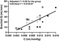
During apnea, MSNA indices were inversely related with vascular compliance, whereas during static handgrip exercise, compliance increased despite increased MSNA.
The elastic components of the forearm vasculature are regulated by adrenergic and myogenic mechanisms during sympatho-excitation, but in a reflex-dependent manner.
Original Research
JAN 2022OlszynskiThe Ultrasound Journal
Performance of an automated ultrasound device in identifying and tracing the heart in porcine cardiac arrest
BACKGROUND: While intra-arrest echocardiography can be used to guide and monitor chest compression quality, it is not currently feasible on the scene of out-of-hospital cardiac arrests. Rapid and automated sonographic localization of the heart may provide first-responders guidance to an optimal area of compression without requiring them to interpret ultrasound images. In this proof-of-concept porcine study, we sought to describe the performance of an automated ultrasound device in correctly identifying and tracing the borders of the heart in three distinct states: pre-arrest, arrest, and late arrest. METHODS: An automated ultrasound device (bladder scanner) was placed on the chests of 7 swine, along the left sternal border (4th-8th intercostal spaces). Scanner-generated images were recorded for each space during pre-arrest, arrest, and finally late arrest. 828 images of the LV and LV outflow tract were randomized and 150 (50/state) selected for analysis. Scanner tracings of the heart were then digitally obscured to facilitate tracing by expert reviewers who were blinded to the physiologic state. Reviewer tracings were compared to bladder scanner tracings; with concordance between these images determined via Sørensen-Dice index (SDI). RESULTS: When compared to human reviewers, the bladder scanner was able to identify and trace the borders during cardiac arrest. The bladder scanner performed best at the time of arrest (SDI 0.900 ± 0.059). As resuscitation efforts continued and time from initial arrest increased, the scanner's performance decreased dramatically (SDI 0.597 ± 0.241 in late arrest). CONCLUSION: An automated ultrasound device (bladder scanner) reliably traced porcine hearts during cardiac arrest. It is possible a device could be developed to indicate where compressions should be performed without requiring the operator to interpret ultrasound images. Further investigation into rapid, automated, sonographic localization of the heart to identify the area of compression in out-of-hospital cardiac arrest is warranted.